
Search Count: 155
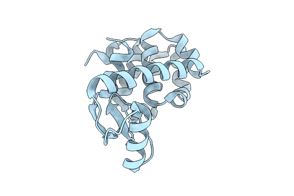 |
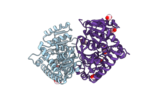
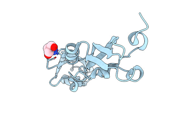
Spitrobot-2 Advances Time-Resolvedcryo-Trapping Crystallography To Under 25 Ms: T4 Lysozyme, Mutant L99A (Apo State)
Organism: Escherichia phage t4
Method: X-RAY DIFFRACTION Release Date: 2025-12-03 Classification: HYDROLASE |
 |
Spitrobot-2 Advances Time-Resolvedcryo-Trapping Crystallography To Under 25 Ms: T4 Lysozyme, Mutant L99A Bound With Indole (1 S Soaking)
Organism: Escherichia phage t4
Method: X-RAY DIFFRACTION Release Date: 2025-12-03 Classification: HYDROLASE Ligands: IND |
 |
Spitrobot-2 Advances Time-Resolvedcryo-Trapping Crystallography To Under 25 Ms: T4 Lysozyme, Mutant L99A Bound With Indole (10 S Soaking)
Organism: Escherichia phage t4
Method: X-RAY DIFFRACTION Release Date: 2025-12-03 Classification: HYDROLASE Ligands: IND, NA |
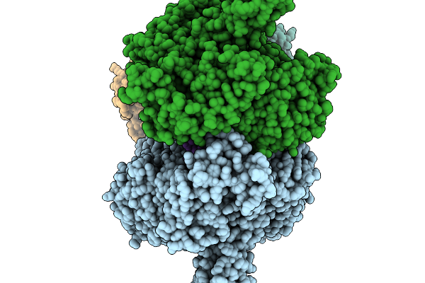 |
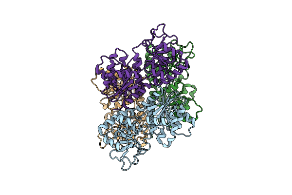
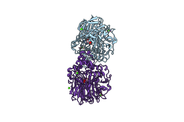
T4 Bacteriophage Replicative Polymerase Captured In Polymerase Exchange State 1
Organism: Escherichia phage t4, Synthetic construct
Method: ELECTRON MICROSCOPY Release Date: 2025-11-19 Classification: replication/DNA |
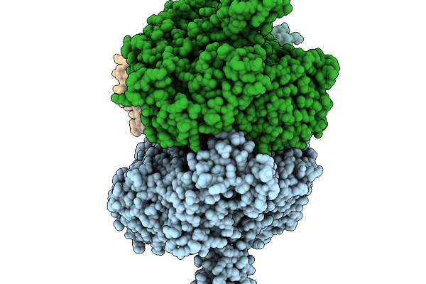 |
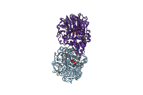
T4 Bacteriophage Replicative Polymerase Captured In Polymerase Exchange State 2
Organism: Escherichia phage t4, Synthetic construct
Method: ELECTRON MICROSCOPY Release Date: 2025-11-19 Classification: replication/DNA |
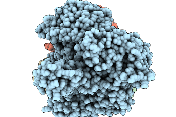 |
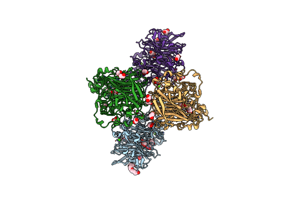
T4 Bacteriophage Replicative Holoenzyme Containing Triple Mutations D75R, Q430E, And K432E In The Exonuclease-Deficient Polymerase
Organism: Escherichia phage t4, Synthetic construct
Method: ELECTRON MICROSCOPY Release Date: 2025-11-19 Classification: Transferase/DNA |
 |
T4 Bacteriophage Replicative Polymerase Captured In Polymerase Exchange State 3
Organism: Escherichia phage t4, Synthetic construct
Method: ELECTRON MICROSCOPY Release Date: 2025-11-19 Classification: Replication/DNA Ligands: CA, D3T |
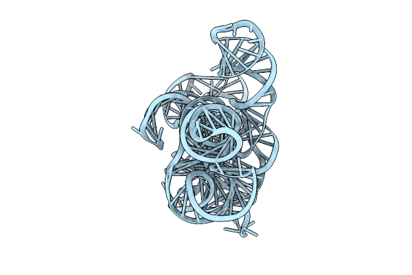 |
Cryo-Em Structure Of Linear Intron Of Thymidylate Synthase (Td) Gene Of Bacteriophage T4
Organism: Escherichia phage t4
Method: ELECTRON MICROSCOPY Release Date: 2025-11-12 Classification: RNA Ligands: MG |
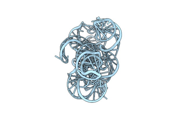 |
Cryo-Em Structure Of Circular Intron Of Thymidylate Synthase (Td) Gene Of Bacteriophage T4
Organism: Escherichia phage t4
Method: ELECTRON MICROSCOPY Release Date: 2025-11-12 Classification: RNA Ligands: MG |
 |
Organism: Candidatus heimdallarchaeota archaeon lc_3, Synthetic construct
Method: ELECTRON MICROSCOPY Release Date: 2025-10-29 Classification: DNA BINDING PROTEIN |
 |
Organism: Candidatus heimdallarchaeota archaeon lc_3, Synthetic construct
Method: ELECTRON MICROSCOPY Release Date: 2025-10-29 Classification: DNA BINDING PROTEIN |
 |
Organism: Candidatus heimdallarchaeota archaeon lc_3, Synthetic construct
Method: ELECTRON MICROSCOPY Release Date: 2025-10-29 Classification: DNA BINDING PROTEIN |
 |
Glycosyltransferase C From The Limosilactobacillus Reuteri Accessory Secretion System. Apo Form.
Organism: Limosilactobacillus reuteri
Method: X-RAY DIFFRACTION Release Date: 2025-04-23 Classification: TRANSFERASE Ligands: CA, GOL |
 |
Glycosyltransferase C From The Limosilactobacillus Reuteri Accessory Secretion System. Complex With Udp.
Organism: Limosilactobacillus reuteri
Method: X-RAY DIFFRACTION Release Date: 2025-04-23 Classification: TRANSFERASE Ligands: UDP |
 |
Crystal Structure L-Lactate Dehydrogenase From Lactobacillus Reuteri In Its Apoform
Organism: Limosilactobacillus reuteri
Method: X-RAY DIFFRACTION Resolution:2.15 Å Release Date: 2025-04-02 Classification: OXIDOREDUCTASE Ligands: EDO |
 |
Structure Of Two Consecutively Arrayed Rib Domains (Rib8-9) From Surface Adhesin Of Limosilactobacillus Reuteri
Organism: Limosilactobacillus reuteri
Method: X-RAY DIFFRACTION Resolution:1.80 Å Release Date: 2025-03-05 Classification: STRUCTURAL PROTEIN Ligands: TRS |
 |
Organism: Limosilactobacillus reuteri
Method: X-RAY DIFFRACTION Release Date: 2025-01-22 Classification: HYDROLASE Ligands: CA, GOL |
 |
Crystal Structure Of Inulosucrase From Lactobacillus Reuteri 121 In Complex With Fructose
Organism: Limosilactobacillus reuteri
Method: X-RAY DIFFRACTION Release Date: 2025-01-22 Classification: HYDROLASE Ligands: FRU, CA |
 |
Crystal Structure Of Inulosucrase From Lactobacillus Reuteri 121 Mutant R544W
Organism: Limosilactobacillus reuteri
Method: X-RAY DIFFRACTION Release Date: 2025-01-22 Classification: HYDROLASE Ligands: CA, EDO, GOL, 1PE, PEG |
 |
Organism: Escherichia coli k-12, Escherichia phage t4
Method: ELECTRON MICROSCOPY Release Date: 2024-11-13 Classification: TRANSCRIPTION/DNA/RNA |