Planned Maintenance: Some services may turn out to be unavailable from 15th January, 2026 to 16th January, 2026. We apologize for the inconvenience!

Planned Maintenance: Some services may turn out to be unavailable from 15th January, 2026 to 16th January, 2026. We apologize for the inconvenience!
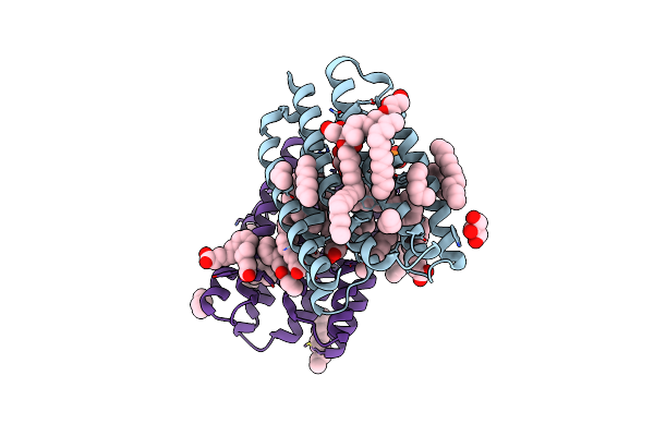 |
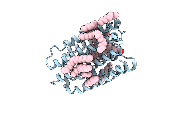
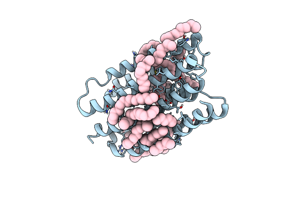
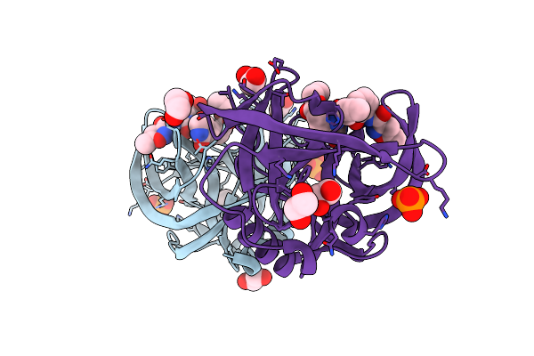
Crystal Structure Of Marine Actinobacteria Clade Rhodopsin (Mar) In The Ground State
Organism: Candidatus actinomarina minuta, Marine actinobacteria clade
Method: X-RAY DIFFRACTION Release Date: 2025-04-02 Classification: MEMBRANE PROTEIN Ligands: OLC, LFA, GOL, RET, PO4 |
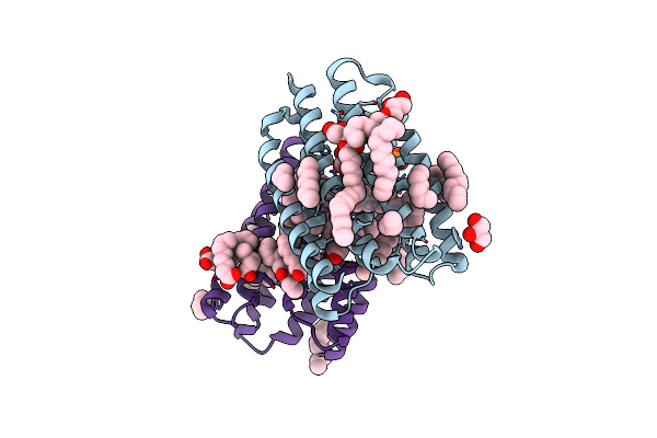 |
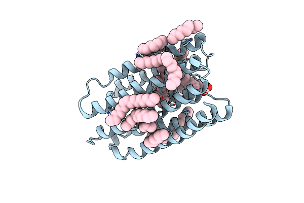
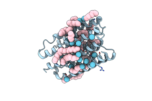
Crystal Structure Of Marine Actinobacteria Clade Rhodopsin (Mar) In The P596 State
Organism: Candidatus actinomarina minuta, Marine actinobacteria clade
Method: X-RAY DIFFRACTION Release Date: 2025-04-02 Classification: MEMBRANE PROTEIN Ligands: OLC, LFA, GOL, RET, PO4 |
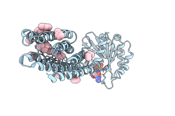 |
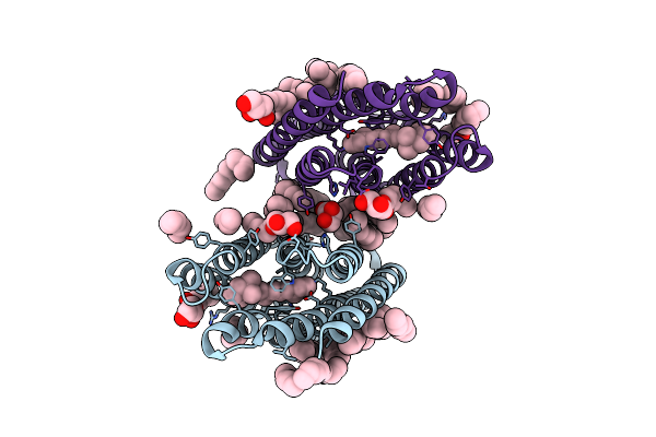
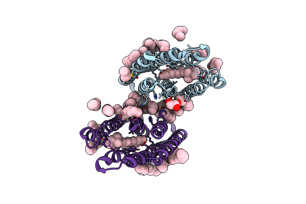
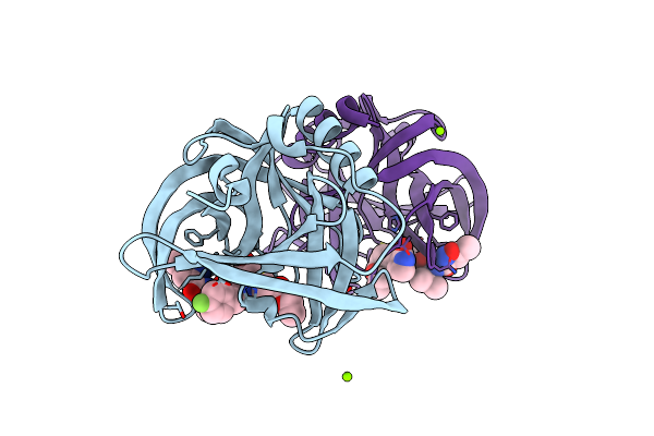
Crystal Structure Of Marine Actinobacteria Clade Rhodopsin (Mar) - Human Gtpase Arf1 (L8K,Q71L) Chimera; Ground State
Organism: Candidatus actinomarina minuta, Homo sapiens, Marine actinobacteria clade
Method: X-RAY DIFFRACTION Release Date: 2025-04-02 Classification: MEMBRANE PROTEIN Ligands: GDP, LFA, RET |
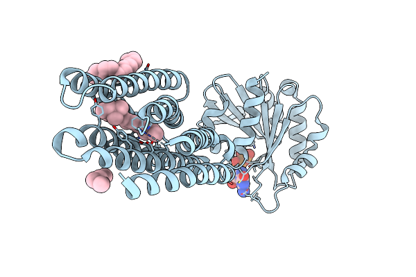 |
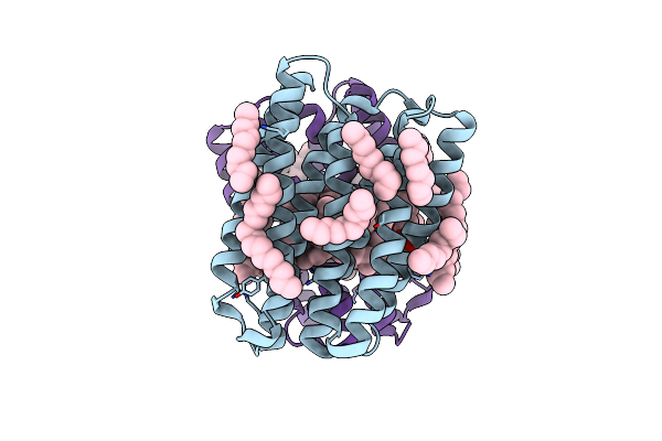
Crystal Structure Of Marine Actinobacteria Clade Rhodopsin (Mar) - Human Gtpase Arf1 (L8K,Q71L) Chimera; N State
Organism: Candidatus actinomarina minuta, Homo sapiens, Marine actinobacteria clade
Method: X-RAY DIFFRACTION Release Date: 2025-04-02 Classification: MEMBRANE PROTEIN Ligands: GDP, LFA, OLA, RET |
 |
Crystal Structure Of Marine Actinobacteria Clade Rhodopsin (Mar) In The O* State
Organism: Candidatus actinomarina minuta, Marine actinobacteria clade
Method: X-RAY DIFFRACTION Release Date: 2025-04-02 Classification: MEMBRANE PROTEIN Ligands: OLA, LFA, RET |
 |
Crystal Structure Of Marine Actinobacteria Clade Rhodopsin (Mar) In The O* State, Ph 8.8
Organism: Candidatus actinomarina minuta, Marine actinobacteria clade
Method: X-RAY DIFFRACTION Release Date: 2025-04-02 Classification: MEMBRANE PROTEIN Ligands: OLA, LFA, RET |
 |
Crystal Structure Of Marine Actinobacteria Clade Rhodopsin (Mar) In The O State Obtained By Cryotrapping
Organism: Candidatus actinomarina minuta, Marine actinobacteria clade
Method: X-RAY DIFFRACTION Release Date: 2025-04-02 Classification: MEMBRANE PROTEIN Ligands: OLA, LFA, RET |
 |
Crystal Structure Of Marine Actinobacteria Clade Rhodopsin (Mar) In The M-Like State
Organism: Candidatus actinomarina minuta
Method: X-RAY DIFFRACTION Resolution:1.60 Å Release Date: 2022-06-01 Classification: MEMBRANE PROTEIN Ligands: LFA, OLB, OLC, RET |
 |
Structure Of Marine Actinobacteria Clade Rhodopsin (Macr) In Orange Form In P1211 Space Group
Organism: Candidatus actinomarina minuta
Method: X-RAY DIFFRACTION Resolution:1.70 Å Release Date: 2022-06-01 Classification: MEMBRANE PROTEIN Ligands: LFA, OLB, OLC |
 |
Crystal Structure Of Marine Actinobacteria Clade Rhodopsin (Mar) In The O State
Organism: Candidatus actinomarina minuta
Method: X-RAY DIFFRACTION Resolution:1.09 Å Release Date: 2022-06-01 Classification: MEMBRANE PROTEIN Ligands: LFA, RET |
 |
Organism: Candidatus actinomarina minuta
Method: X-RAY DIFFRACTION Resolution:2.25 Å Release Date: 2022-04-27 Classification: MEMBRANE PROTEIN Ligands: RET, LFA, HEX, KR |
 |
Organism: Cocos nucifera
Method: X-RAY DIFFRACTION Resolution:1.85 Å Release Date: 2017-10-25 Classification: ALLERGEN |
 |
Organism: Candidatus actinomarina minuta
Method: X-RAY DIFFRACTION Resolution:2.00 Å Release Date: 2017-05-31 Classification: UNKNOWN FUNCTION Ligands: LFA, IOD, OLC, RET |
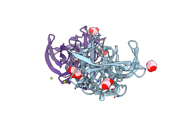 |
Organism: Human enterovirus b
Method: X-RAY DIFFRACTION Resolution:1.90 Å Release Date: 2011-09-07 Classification: HYDROLASE Ligands: GOL, MG |
 |
Organism: Human enterovirus b
Method: X-RAY DIFFRACTION Resolution:1.32 Å Release Date: 2011-09-07 Classification: HYDROLASE/HYDROLASE INHIBITOR Ligands: XNV, EDO, PI, NH4 |
 |
Organism: Human enterovirus b
Method: X-RAY DIFFRACTION Resolution:1.50 Å Release Date: 2011-09-07 Classification: HYDROLASE/HYDROLASE INHIBITOR Ligands: AG7, MG, CL |
 |
 |
Organism: Steromapedaliodes albonotata australis
Method: Alphafold Release Date: Classification: NA Ligands: NA |
 |
Organism: Steromapedaliodes albonotata josefinae
Method: Alphafold Release Date: Classification: NA Ligands: NA |
 |