Planned Maintenance: Some services may turn out to be unavailable from 15th January, 2026 to 16th January, 2026. We apologize for the inconvenience!

Planned Maintenance: Some services may turn out to be unavailable from 15th January, 2026 to 16th January, 2026. We apologize for the inconvenience!
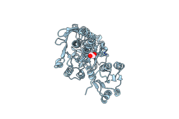 |
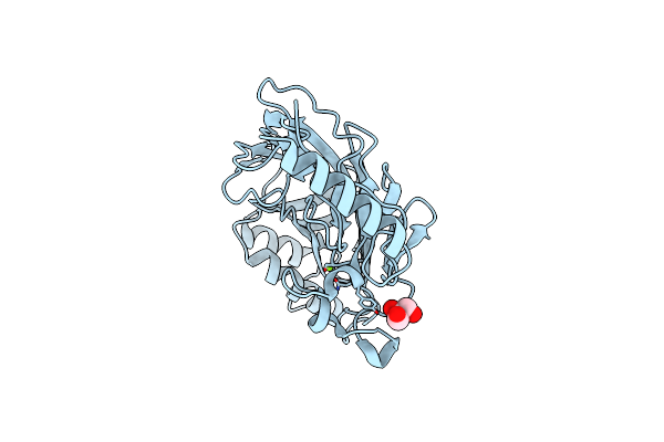
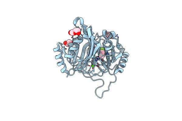
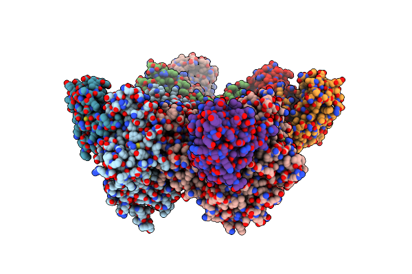
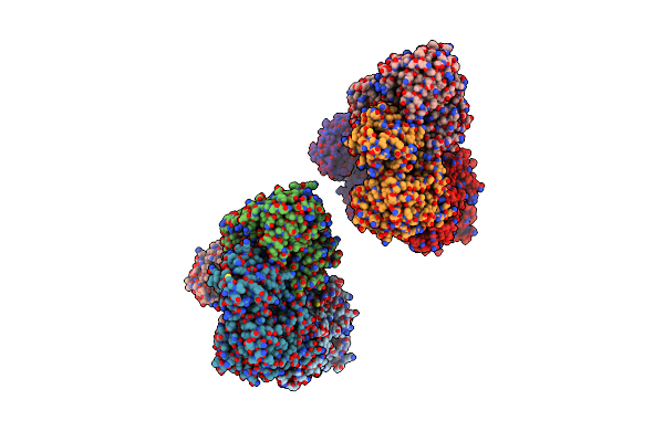
Crystal Structure Of Apo-Form Udp-N-Acetylmuramic Acid L-Alanine Ligase (Murc) From Roseburia Faecis
Organism: Roseburia faecis
Method: X-RAY DIFFRACTION Release Date: 2025-04-09 Classification: LIGASE Ligands: EDO |
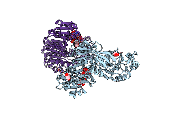 |
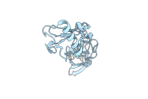
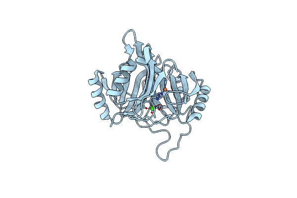
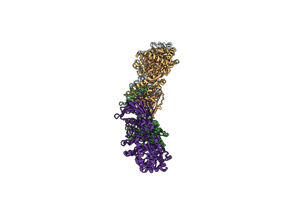
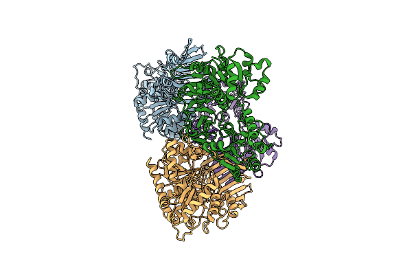
Crystal Structure Of Udp-N-Acetylmuramic Acid L-Alanine Ligase (Murc) From Roseburia Faecis In Complex With Unam
Organism: Roseburia faecis
Method: X-RAY DIFFRACTION Release Date: 2025-04-09 Classification: LIGASE Ligands: EDO, EPZ |
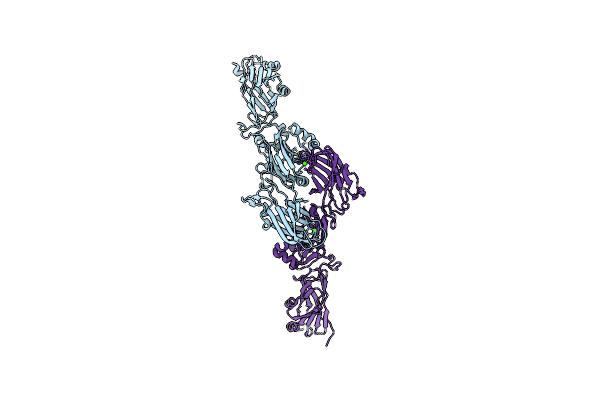 |
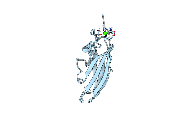
Organism: Streptococcus gordonii
Method: X-RAY DIFFRACTION Resolution:2.70 Å Release Date: 2021-09-08 Classification: CELL ADHESION Ligands: CA |
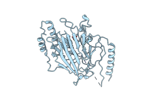 |
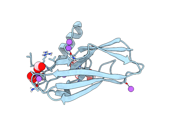
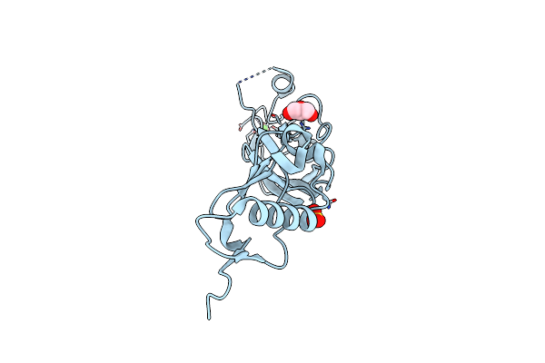
The Structure Of The Streptococcus Gordonii Surface Protein Sspb In Complex With Tev Peptide Provides Clues To The Adherence Of Oral Streptococcal Adherence To Salivary Agglutinin
Organism: Streptococcus gordonii
Method: X-RAY DIFFRACTION Resolution:2.00 Å Release Date: 2020-08-12 Classification: CELL ADHESION Ligands: CA |
 |
The Structure Of The Streptococcus Gordonii Surface Protein Sspb In Complex With Tev Peptide Provides Clues To The Adherence Of Oral Streptococcal Adherence To Salivary Agglutinin
Organism: Streptococcus gordonii
Method: X-RAY DIFFRACTION Resolution:1.80 Å Release Date: 2020-08-12 Classification: CELL ADHESION Ligands: MG, GOL |
 |
Organism: Streptococcus gordonii
Method: X-RAY DIFFRACTION Resolution:1.03 Å Release Date: 2020-02-19 Classification: SUGAR BINDING PROTEIN Ligands: CA |
 |
Organism: Streptococcus gordonii
Method: X-RAY DIFFRACTION Resolution:1.30 Å Release Date: 2020-02-19 Classification: SUGAR BINDING PROTEIN |
 |
Organism: Streptococcus gordonii
Method: X-RAY DIFFRACTION Resolution:1.60 Å Release Date: 2020-02-19 Classification: SUGAR BINDING PROTEIN Ligands: CA |
 |
Organism: Streptococcus gordonii
Method: X-RAY DIFFRACTION Resolution:1.90 Å Release Date: 2020-02-19 Classification: SUGAR BINDING PROTEIN Ligands: GOL, CA, SO4 |
 |
Organism: Diaphorobacter sp. ds2
Method: X-RAY DIFFRACTION Resolution:2.20 Å Release Date: 2019-06-12 Classification: OXIDOREDUCTASE Ligands: CA, FE, 3FA, PEG, EDO |
 |
Organism: Diaphorobacter sp. ds2
Method: X-RAY DIFFRACTION Resolution:2.40 Å Release Date: 2019-05-22 Classification: OXIDOREDUCTASE Ligands: CA, FE |
 |
Organism: Diaphorobacter sp. ds2
Method: X-RAY DIFFRACTION Resolution:2.40 Å Release Date: 2019-04-17 Classification: OXIDOREDUCTASE Ligands: FE, MCT, PEG, EDO, CA |
 |
Organism: Streptococcus gordonii
Method: X-RAY DIFFRACTION Resolution:3.11 Å Release Date: 2018-02-21 Classification: CELL ADHESION |
 |
Organism: Streptococcus gordonii
Method: X-RAY DIFFRACTION Resolution:2.77 Å Release Date: 2018-02-21 Classification: CELL ADHESION |
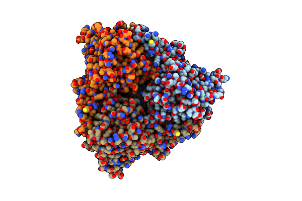 |
Oxygenase Component Of 3-Nitrotoluene Dioxygenase From Diaphorobacter Sp. Strain Ds2
Organism: Diaphorobacter sp. ds2
Method: X-RAY DIFFRACTION Resolution:2.90 Å Release Date: 2017-04-19 Classification: METAL BINDING PROTEIN Ligands: FE, FES |
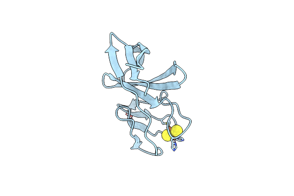 |
Ferredoxin Component Of 3-Nitrotoluene Dioxygenase From Diaphorobacter Sp. Strain Ds2
Organism: Diaphorobacter sp. ds2
Method: X-RAY DIFFRACTION Resolution:2.40 Å Release Date: 2016-07-27 Classification: METAL BINDING PROTEIN Ligands: FES, FE |
 |
Organism: Streptococcus gordonii
Method: X-RAY DIFFRACTION Resolution:1.25 Å Release Date: 2016-04-13 Classification: SUGAR BINDING PROTEIN Ligands: MG |
 |
Organism: Streptococcus gordonii
Method: X-RAY DIFFRACTION Resolution:2.92 Å Release Date: 2016-03-02 Classification: TRANSFERASE/CHAPERONE Ligands: MG |
 |
Organism: Streptococcus gordonii
Method: X-RAY DIFFRACTION Resolution:3.84 Å Release Date: 2016-03-02 Classification: TRANSFERASE/CHAPERONE Ligands: UDP, NAG, MG |
 |
Organism: Streptococcus gordonii
Method: X-RAY DIFFRACTION Resolution:1.40 Å Release Date: 2011-08-10 Classification: SUGAR BINDING PROTEIN Ligands: K, GOL, HDZ, NA |