
Search Count: 41
 |
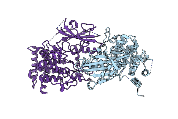
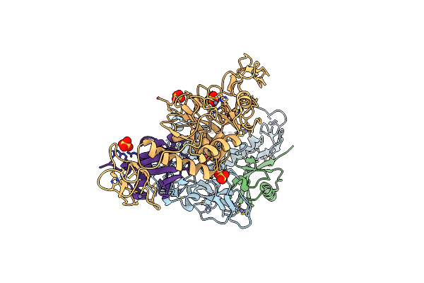
Structure Of The Ligand Binding Domain Of The Chemoreceptor Mkca (Dsomk10_Rs0100305) Of Dickeya Solani Mk10 In Complex With Choline
Organism: Dickeya solani mk10
Method: X-RAY DIFFRACTION Release Date: 2025-08-06 Classification: SIGNALING PROTEIN Ligands: CHT, GOL, EDO, SO4 |
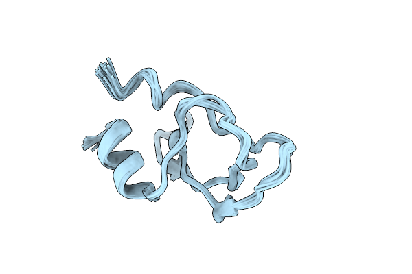 |
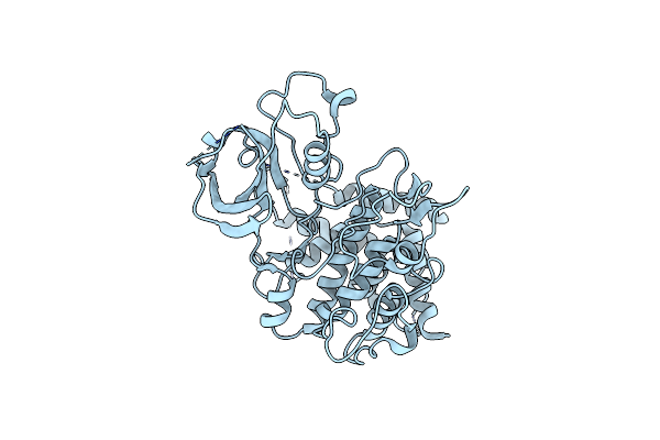
Serine Protease Inhibitor Hciq2C1 From Heteractis Crispa With Trpa1 Mediated Antinociceptive Activity
|
 |
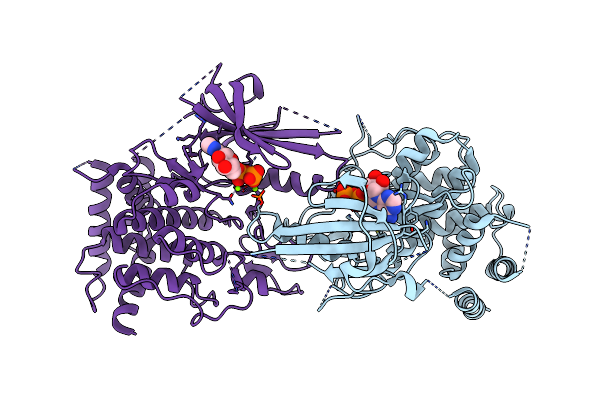
Organism: Pediculus humanus corporis
Method: ELECTRON MICROSCOPY Release Date: 2024-01-31 Classification: TRANSFERASE |
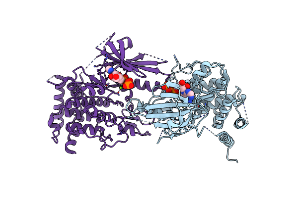 |
Organism: Pediculus humanus corporis
Method: ELECTRON MICROSCOPY Release Date: 2024-01-31 Classification: TRANSFERASE Ligands: ANP, MG |
 |
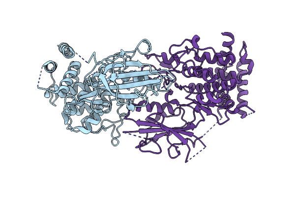
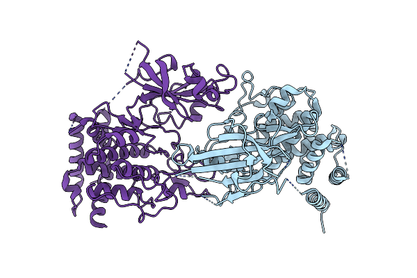
Structure Of Adp-Bound And Phosphorylated Pediculus Humanus (Ph) Pink1 Dimer
Organism: Pediculus humanus corporis
Method: ELECTRON MICROSCOPY Release Date: 2024-01-31 Classification: TRANSFERASE Ligands: ADP, MG |
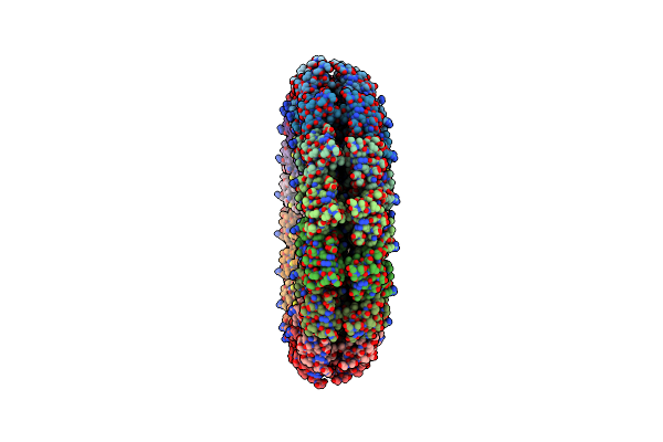 |
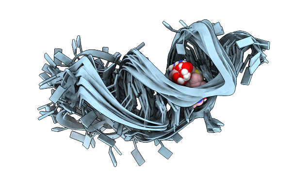
An Asymmetric Disk Assembly Formed By Tandem Dimers Of The Tobacco Mosaic Viral Capsid Protein (Tmv)
Organism: Tobacco mosaic virus (vulgare)
Method: X-RAY DIFFRACTION Resolution:2.80 Å Release Date: 2023-03-01 Classification: VIRAL PROTEIN |
 |
Organism: Methylococcus capsulatus str. texas = atcc 19069
Method: X-RAY DIFFRACTION Resolution:1.60 Å Release Date: 2022-12-14 Classification: TRANSPORT PROTEIN Ligands: CL |
 |
Organism: Methylococcus capsulatus str. texas = atcc 19069
Method: X-RAY DIFFRACTION Resolution:1.91 Å Release Date: 2022-12-14 Classification: TRANSPORT PROTEIN Ligands: PEG, CIT |
 |
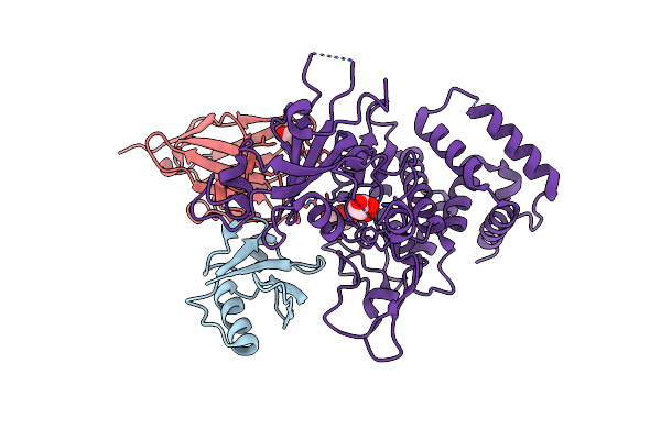
Crystal Structure Of Peptono Toxin, A Diphtheria Toxin Homolog, From Seinonella Peptonophila
Organism: Seinonella peptonophila
Method: X-RAY DIFFRACTION Resolution:2.19 Å Release Date: 2022-04-20 Classification: TOXIN Ligands: IOD |
 |
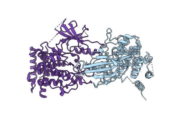
Structure Of Dimeric Phosphorylated Pediculus Humanus (Ph) Pink1 With Kinked Alpha-C Helix In Chain B
Organism: Pediculus humanus corporis
Method: ELECTRON MICROSCOPY Release Date: 2022-01-12 Classification: TRANSFERASE |
 |
Structure Of Dimeric Phosphorylated Pediculus Humanus (Ph) Pink1 With Extended Alpha-C Helix In Chain B
Organism: Pediculus humanus corporis
Method: ELECTRON MICROSCOPY Release Date: 2022-01-12 Classification: TRANSFERASE |
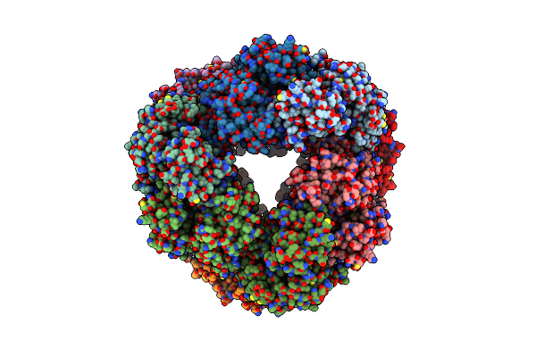 |
Structure Of Dodecameric Unphosphorylated Pediculus Humanus (Ph) Pink1 D357A Mutant
Organism: Pediculus humanus corporis
Method: ELECTRON MICROSCOPY Release Date: 2022-01-12 Classification: TRANSFERASE |
 |
Structure Of Dimeric Unphosphorylated Pediculus Humanus (Ph) Pink1 D357A Mutant
Organism: Pediculus humanus corporis
Method: ELECTRON MICROSCOPY Release Date: 2022-01-12 Classification: TRANSFERASE |
 |
Organism: Pediculus humanus corporis
Method: X-RAY DIFFRACTION Resolution:3.53 Å Release Date: 2021-12-22 Classification: TRANSFERASE |
 |
A Circular Permutant Of The Tobacco Mosaic Virus (Tmv) Mutant Q101H Coordinated With Heme
Organism: Tobacco mosaic virus (strain vulgare)
Method: X-RAY DIFFRACTION Resolution:3.00 Å Release Date: 2020-12-09 Classification: VIRAL PROTEIN Ligands: HEM |
 |
Organism: Tobacco mosaic virus (strain vulgare)
Method: X-RAY DIFFRACTION Resolution:3.00 Å Release Date: 2020-12-09 Classification: VIRAL PROTEIN |
 |
Organism: Proteobacteria
Method: SOLUTION NMR Release Date: 2019-01-09 Classification: RNA Ligands: SAH |
 |
Organism: Homo sapiens, Lama glama, Pediculus humanus corporis
Method: X-RAY DIFFRACTION Resolution:3.10 Å Release Date: 2017-11-08 Classification: TRANSFERASE Ligands: GOL |
 |
Novel Inter-Subunit Contacts In Barley Stripe Mosaic Virus Revealed By Cryo-Em
Organism: Barley stripe mosaic virus, Tobacco mosaic virus
Method: ELECTRON MICROSCOPY Resolution:4.10 Å Release Date: 2015-09-02 Classification: VIRUS |
 |
Organism: Pediculus humanus subsp. corporis, Homo sapiens
Method: X-RAY DIFFRACTION Resolution:2.62 Å Release Date: 2015-07-22 Classification: SIGNALING PROTEIN Ligands: ZN, SO4 |