
Search Count: 31
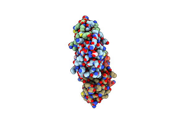 |
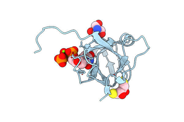
Mason-Pfizer Monkey Virus Protease Mutant C7A/D26N/C106A In Complex With Peptidomimetic Inhibitor
Organism: Mason-pfizer monkey virus, Synthetic construct
Method: X-RAY DIFFRACTION Resolution:1.93 Å Release Date: 2021-12-15 Classification: HYDROLASE Ligands: ACT, 1PE |
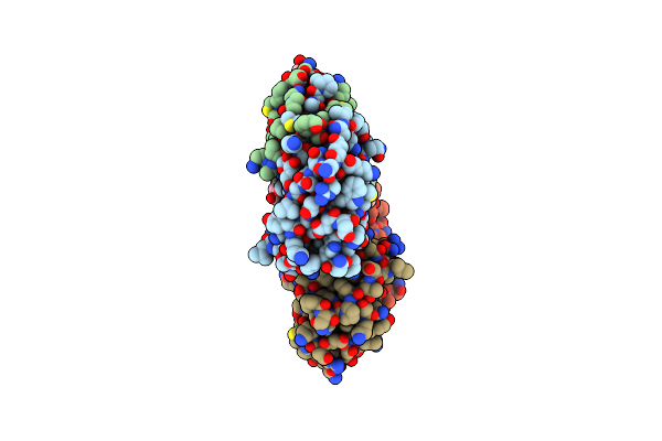 |
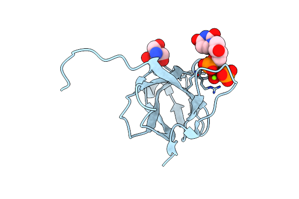
Mason-Pfizer Monkey Virus Protease Mutant C7A/D26N/C106A In Complex With Peptidomimetic Inhibitor
Organism: Mason-pfizer monkey virus, Synthetic construct
Method: X-RAY DIFFRACTION Resolution:2.43 Å Release Date: 2021-12-15 Classification: HYDROLASE Ligands: 1PE |
 |
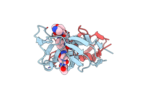
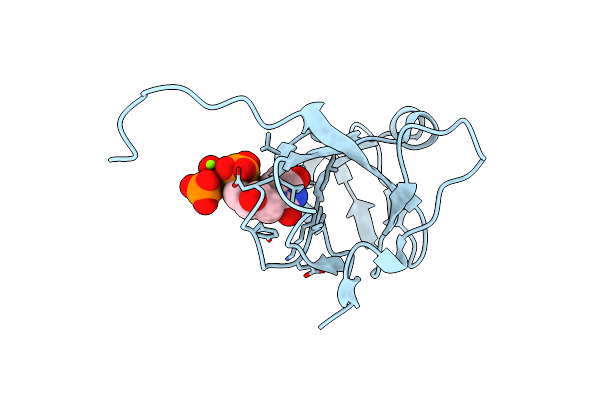
Crystal Structure Of Dimeric M-Pmv Protease C7A/D26N/C106A Mutant In Complex With Inhibitor
Organism: Mason-pfizer monkey virus, Synthetic construct
Method: X-RAY DIFFRACTION Resolution:1.90 Å Release Date: 2019-10-16 Classification: HYDROLASE |
 |
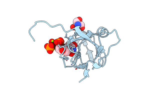
Crystal Structure Of Dimeric M-Pmv Protease D26N Mutant In Complex With Inhibitor
Organism: Mason-pfizer monkey virus, Synthetic construct
Method: X-RAY DIFFRACTION Resolution:1.64 Å Release Date: 2019-10-16 Classification: HYDROLASE |
 |
Organism: Mason-pfizer monkey virus
Method: X-RAY DIFFRACTION Resolution:1.98 Å Release Date: 2019-10-16 Classification: HYDROLASE |
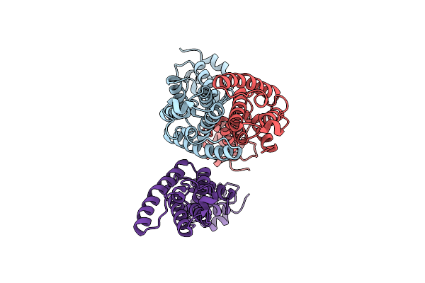 |
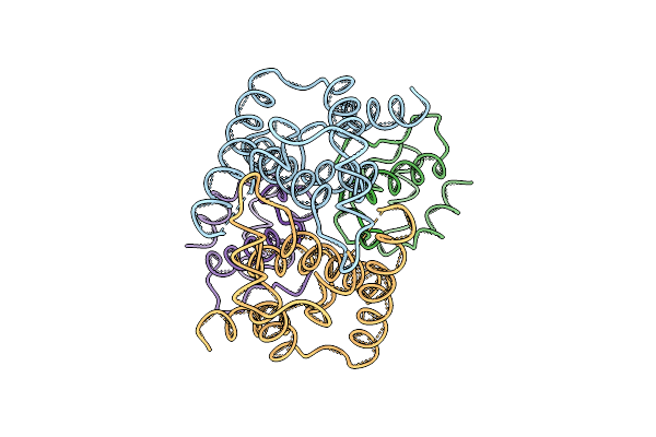
Organism: Mason-pfizer monkey virus
Method: ELECTRON MICROSCOPY Release Date: 2018-12-05 Classification: VIRAL PROTEIN |
 |
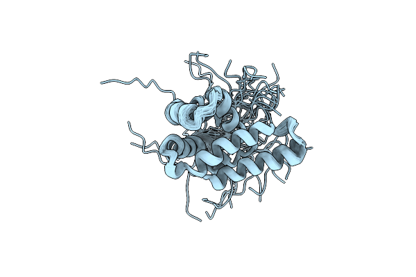
Organism: Mason-pfizer monkey virus
Method: SOLUTION NMR Release Date: 2016-10-05 Classification: VIRAL PROTEIN |
 |
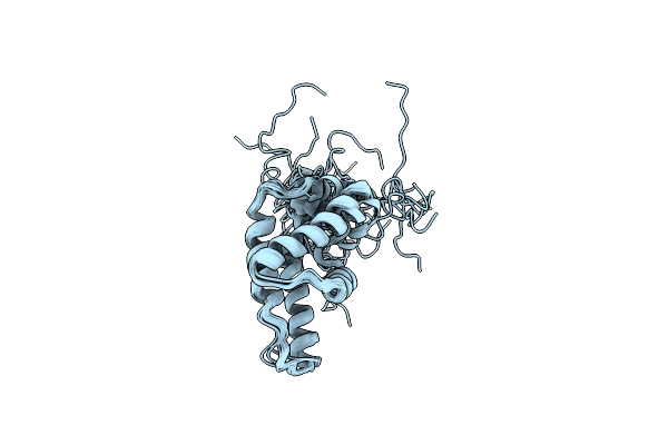
Organism: Mason-pfizer monkey virus
Method: SOLUTION NMR Release Date: 2016-07-27 Classification: VIRAL PROTEIN Ligands: MYR |
 |
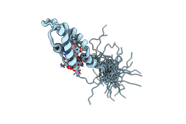
Solution Structure Of Myristoylated Y28F/Y67F Mutant Of The Mason-Pfizer Monkey Virus Matrix Protein
Organism: Mason-pfizer monkey virus
Method: SOLUTION NMR Release Date: 2015-04-01 Classification: VIRAL PROTEIN |
 |
Organism: Mason-pfizer monkey virus
Method: X-RAY DIFFRACTION Resolution:1.70 Å Release Date: 2013-10-23 Classification: VIRAL PROTEIN Ligands: CL |
 |
Structure Of The Immature Retroviral Capsid At 8A Resolution By Cryo- Electron Microscopy
Organism: Mason-pfizer monkey virus
Method: ELECTRON MICROSCOPY Resolution:7.00 Å Release Date: 2012-05-30 Classification: VIRAL PROTEIN |
 |
Structure Of The Immature Retroviral Capsid At 8A Resolution By Cryo- Electron Microscopy
Organism: Mason-pfizer monkey virus
Method: ELECTRON MICROSCOPY Resolution:7.00 Å Release Date: 2012-05-30 Classification: VIRAL PROTEIN |
 |
Organism: Mason-pfizer monkey virus
Method: X-RAY DIFFRACTION Resolution:1.65 Å Release Date: 2011-10-12 Classification: HYDROLASE Ligands: DUP, MG, TRS, DTT |
 |
Organism: Mason-pfizer monkey virus
Method: X-RAY DIFFRACTION Resolution:1.85 Å Release Date: 2011-10-12 Classification: HYDROLASE Ligands: MG, DUP, TRS |
 |
Crystal Structure Of M-Pmv Dutpase With A Mixed Population Of Substrate (Dupnpp) And Post-Inversion Product (Dump) In The Active Sites
Organism: Mason-pfizer monkey virus
Method: X-RAY DIFFRACTION Resolution:1.75 Å Release Date: 2011-10-12 Classification: HYDROLASE Ligands: MG, TRS, DUP, UMP |
 |
Crystal Structure Of M-Pmv Dutpase With A Mixed Population Of Substrate (Dupnpp) And Post-Inversion Product (Dump) In The Active Sites
Organism: Mason-pfizer monkey virus
Method: X-RAY DIFFRACTION Resolution:1.85 Å Release Date: 2011-10-12 Classification: HYDROLASE Ligands: UMP, DUP, MG |
 |
Crystal Structure Of M-Pmv Dutpase With A Mixed Population Of Substrate (Dupnpp) And Post-Inversion Product (Dump) In The Active Sites
Organism: Mason-pfizer monkey virus
Method: X-RAY DIFFRACTION Resolution:1.60 Å Release Date: 2011-10-12 Classification: HYDROLASE Ligands: UMP, MG, DUP, TRS |
 |
Organism: Mason-pfizer monkey virus
Method: X-RAY DIFFRACTION Resolution:1.40 Å Release Date: 2011-10-12 Classification: HYDROLASE Ligands: UMP, TRS |
 |
Organism: Mason-pfizer monkey virus
Method: X-RAY DIFFRACTION Resolution:1.80 Å Release Date: 2011-10-12 Classification: HYDROLASE Ligands: UMP |
 |
Organism: Mason-pfizer monkey virus
Method: X-RAY DIFFRACTION Resolution:1.83 Å Release Date: 2011-10-12 Classification: HYDROLASE Ligands: UMP |