
Search Count: 49
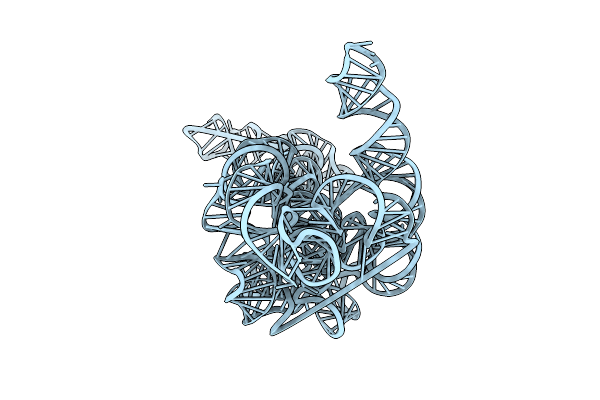 |
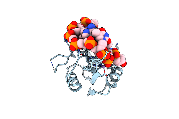
Organism: Powassan virus
Method: ELECTRON MICROSCOPY Release Date: 2025-05-28 Classification: RNA Ligands: MG |
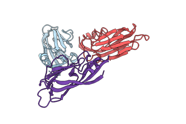 |
Cryoem Structure Of Modified Turnip Yellows Virus Devoid Of Minor Capsid Protein Readthrough Domain
Organism: Turnip yellows virus
Method: ELECTRON MICROSCOPY Release Date: 2025-05-07 Classification: VIRUS |
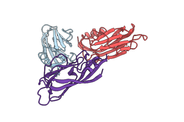 |
Organism: Turnip yellows virus
Method: ELECTRON MICROSCOPY Release Date: 2024-06-05 Classification: VIRUS |
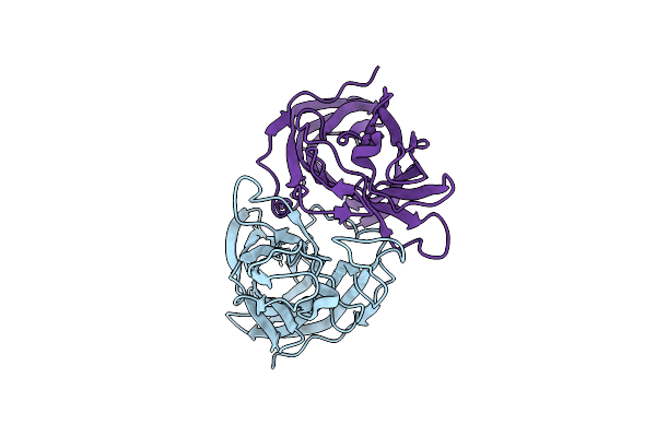 |
Organism: Turnip yellows virus
Method: X-RAY DIFFRACTION Resolution:1.53 Å Release Date: 2022-11-02 Classification: VIRAL PROTEIN |
 |
Organism: Tomato yellow leaf curl virus, Tomato yellow leaf curl virus-il [mx: slp:11]
Method: X-RAY DIFFRACTION Resolution:2.04 Å Release Date: 2022-09-21 Classification: VIRAL PROTEIN Ligands: MN |
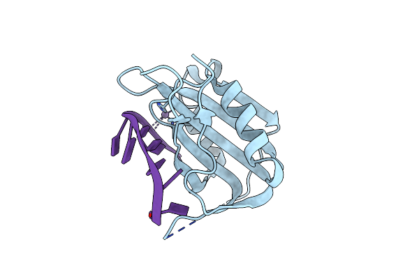 |
Wheat Dwarf Virus Rep Domain Circular Permutation Complexed With A Single-Stranded Dna 10-Mer Comprising The Cleavage Site And Mn2+
Organism: Wheat dwarf virus
Method: X-RAY DIFFRACTION Resolution:1.73 Å Release Date: 2021-11-17 Classification: REPLICATION/DNA Ligands: MN |
 |
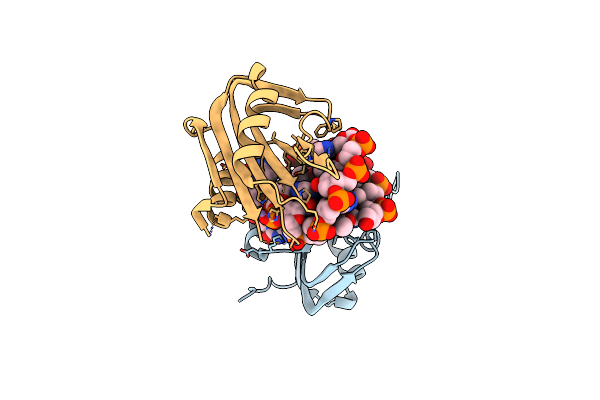
Wheat Dwarf Virus Rep Domain Complexed With A Single-Stranded Dna 10-Mer Comprising The Cleavage Site
Organism: Wheat dwarf virus
Method: X-RAY DIFFRACTION Resolution:1.80 Å Release Date: 2020-12-16 Classification: REPLICATION/DNA Ligands: MN |
 |
Wheat Dwarf Virus Rep Domain Complexed With A Single-Stranded Dna 8-Mer Comprising The Cleavage Site
Organism: Wheat dwarf virus
Method: X-RAY DIFFRACTION Resolution:2.61 Å Release Date: 2020-12-16 Classification: REPLICATION/DNA Ligands: MN |
 |
Organism: Wheat dwarf virus
Method: X-RAY DIFFRACTION Resolution:1.24 Å Release Date: 2019-12-11 Classification: REPLICATION Ligands: GOL |
 |
Hexagonal Form Of Phosphopantetheine Adenylyltransferase From Mycobacterium Tuberculosis
Organism: Mycobacterium tuberculosis bt1
Method: X-RAY DIFFRACTION Resolution:2.00 Å Release Date: 2014-08-13 Classification: TRANSFERASE Ligands: SO4 |
 |
Crystal Structure Of Amidohydrolase Map2389C (Target Efi-500390) From Mycobacterium Avium Subsp. Paratuberculosis K-10
Organism: Mycobacterium avium subsp. paratuberculosis s397
Method: X-RAY DIFFRACTION Resolution:1.60 Å Release Date: 2012-03-21 Classification: HYDROLASE Ligands: CD, CL, MG, SO4 |
 |
Organism: Macrophoma commelinae
Method: X-RAY DIFFRACTION Resolution:1.70 Å Release Date: 2003-04-01 Classification: LYASE Ligands: MG, PYR |
 |
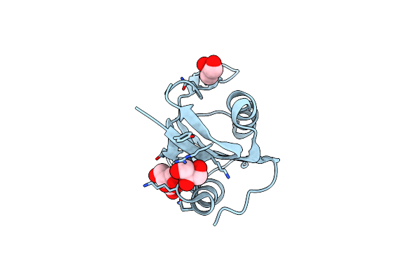
Structure Of Ferricytochrome C(Prime) From Rhodospirillum Molischianum At 1.67 Angstroms Resolution
Organism: Phaeospirillum molischianum
Method: X-RAY DIFFRACTION Resolution:1.67 Å Release Date: 1986-01-21 Classification: ELECTRON TRANSPORT (HEME PROTEIN) Ligands: HEC |
 |
 |
 |
 |
 |
 |
 |