Planned Maintenance: Some services may turn out to be unavailable from 15th January, 2026 to 16th January, 2026. We apologize for the inconvenience!

Planned Maintenance: Some services may turn out to be unavailable from 15th January, 2026 to 16th January, 2026. We apologize for the inconvenience!
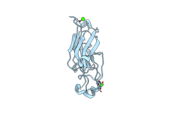 |
Crystal Structure Of The Receptor Binding Domain Of The Spike Protein P1 From Bacteriophage Pm2
Organism: Pseudoalteromonas phage pm2
Method: X-RAY DIFFRACTION Resolution:2.26 Å Release Date: 2008-09-16 Classification: VIRAL PROTEIN Ligands: CA |
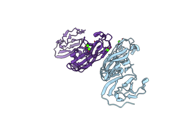 |
Crystal Structure Of The Stem And Receptor Binding Domain Of The Spike Protein P1 From Bacteriophage Pm2
Organism: Pseudoalteromonas phage pm2
Method: X-RAY DIFFRACTION Resolution:1.77 Å Release Date: 2008-09-16 Classification: VIRAL PROTEIN Ligands: CA, CL |
 |
 |
 |
NA
|
 |
NA
|
 |
NA
Organism: Lepidoptera sp. BOLD:AAU7518
Method: Alphafold Release Date: Classification: NA Ligands: NA |