
Search Count: 26
 |
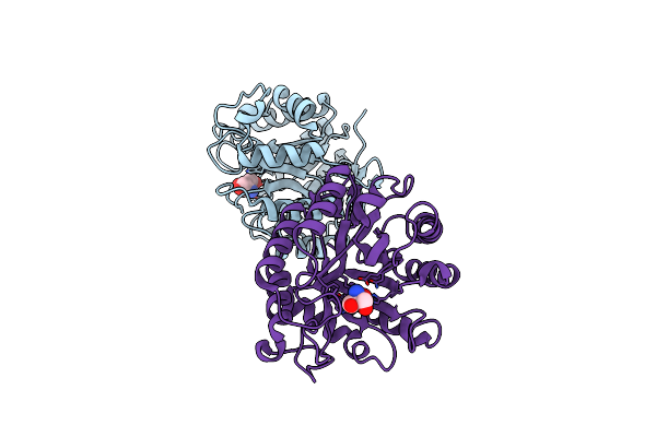
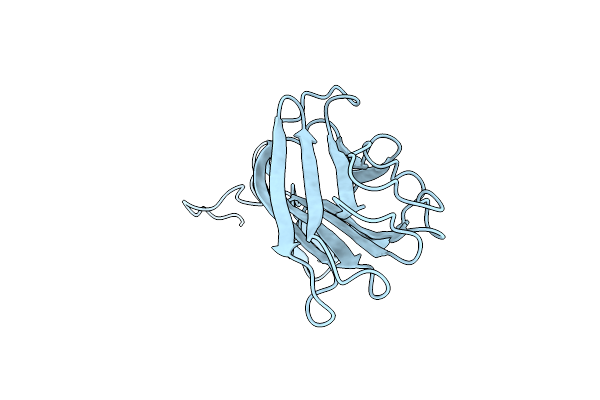
Organism: Enterobacteria phage prd1
Method: X-RAY DIFFRACTION Resolution:1.70 Å Release Date: 2025-04-09 Classification: DNA BINDING PROTEIN |
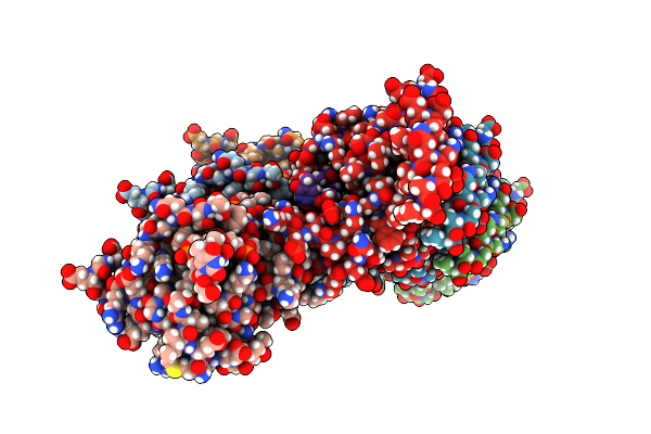 |
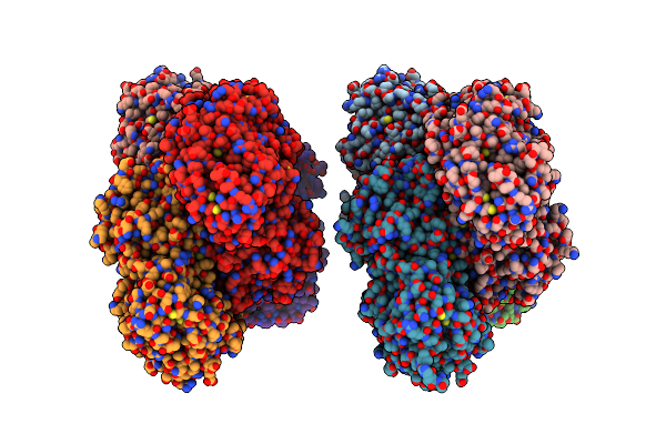
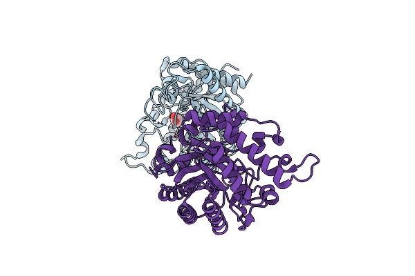
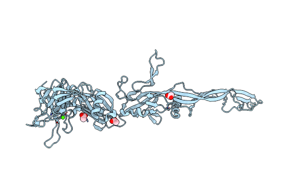
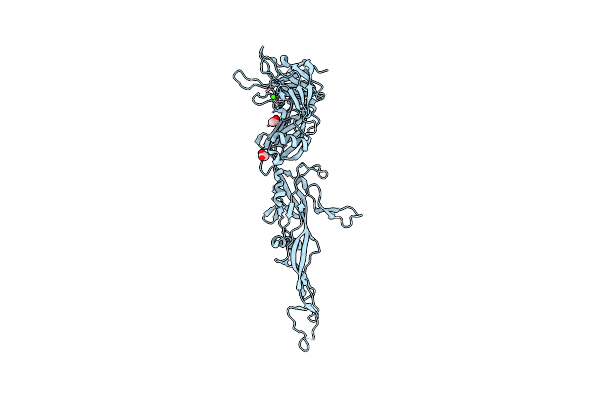
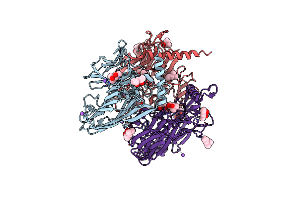
Enterobacteriaphage Prd1 - P12 Protein Filament In Complex With Poly(Dt) Ssdna
Organism: Enterobacteria phage prd1, Synthetic construct
Method: ELECTRON MICROSCOPY Resolution:2.75 Å Release Date: 2025-03-19 Classification: REPLICATION |
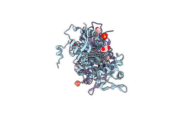 |
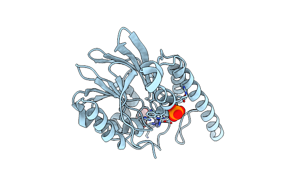
Organism: Soil metagenome
Method: X-RAY DIFFRACTION Resolution:1.73 Å Release Date: 2024-12-25 Classification: HYDROLASE Ligands: ACT, PEG, 1PE, SO4 |
 |
Organism: Soil metagenome
Method: X-RAY DIFFRACTION Resolution:1.80 Å Release Date: 2019-04-10 Classification: HYDROLASE Ligands: GOL |
 |
Crystal Structure Of Gh31 Alpha-Xylosidase From A Soil Metagenome Complexed With Xylose
Organism: Soil metagenome
Method: X-RAY DIFFRACTION Resolution:2.10 Å Release Date: 2019-04-10 Classification: HYDROLASE Ligands: XYS |
 |
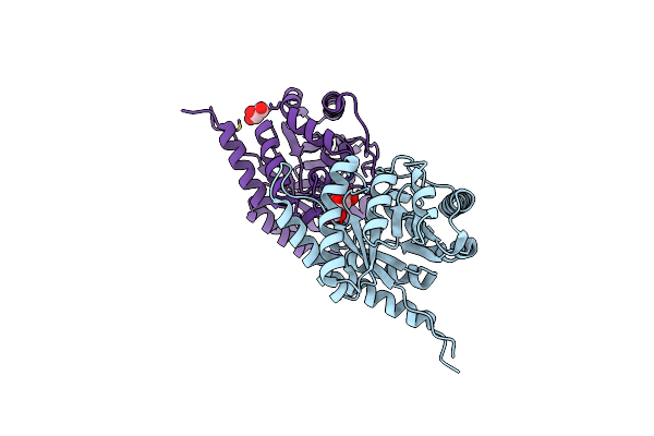
Crystal Structure Of Mem-A1, A Subclass B3 Metallo-Beta-Lactamase Isolated From A Soil Metagenome Library
Organism: Soil metagenome
Method: X-RAY DIFFRACTION Resolution:1.78 Å Release Date: 2018-07-25 Classification: HYDROLASE Ligands: ZN, PO4, GOL |
 |
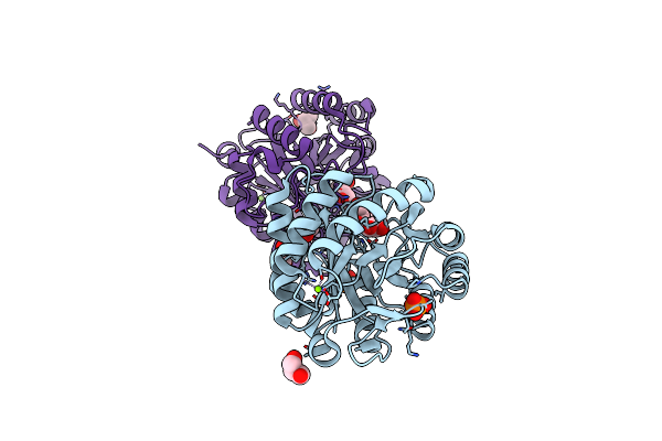
1.73 Angstrom Resolution Crystal Structure Of Dihydropteroate Synthase (Folp-Smz_B27) From Soil Uncultured Bacterium.
Organism: Soil metagenome
Method: X-RAY DIFFRACTION Resolution:1.73 Å Release Date: 2017-05-03 Classification: HYDROLASE,OXIDOREDUCTASE Ligands: MLA, CL, K, TAR, GOL |
 |
Organism: Soil metagenome
Method: X-RAY DIFFRACTION Resolution:2.20 Å Release Date: 2017-01-18 Classification: HYDROLASE Ligands: GOL, MG, PO4 |
 |
Organism: Soil metagenome
Method: X-RAY DIFFRACTION Resolution:1.80 Å Release Date: 2014-02-05 Classification: HYDROLASE Ligands: TRS |
 |
Structure Of A Novel Gh10 Endoxylanase Retrieved From Sugarcane Soil Metagenome
Organism: Soil metagenome
Method: X-RAY DIFFRACTION Resolution:2.74 Å Release Date: 2013-10-23 Classification: HYDROLASE Ligands: GOL |
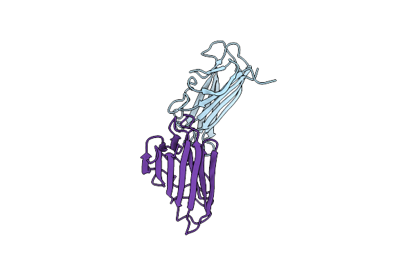 |
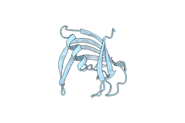
Organism: Enterobacteria phage prd1
Method: X-RAY DIFFRACTION Resolution:2.00 Å Release Date: 2005-04-26 Classification: VIRAL PROTEIN |
 |
Organism: Enterobacteria phage prd1
Method: X-RAY DIFFRACTION Resolution:2.40 Å Release Date: 2005-04-26 Classification: VIRAL PROTEIN |
 |
Organism: Enterobacteria phage prd1
Method: X-RAY DIFFRACTION Resolution:2.60 Å Release Date: 2005-04-26 Classification: VIRAL PROTEIN |
 |
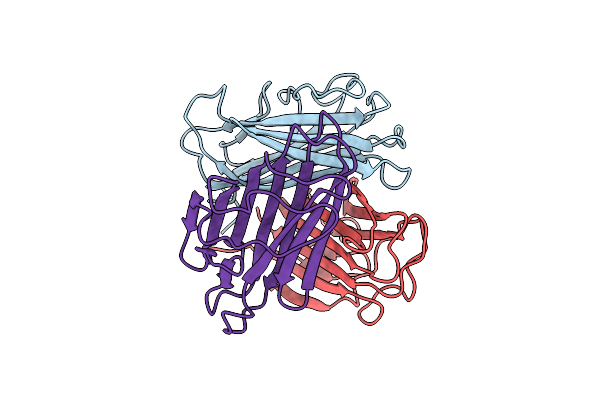
Organism: Enterobacteria phage prd1
Method: X-RAY DIFFRACTION Resolution:2.40 Å Release Date: 2003-04-08 Classification: VIRAL PROTEIN Ligands: ACT, CA |
 |
Organism: Enterobacteria phage prd1
Method: X-RAY DIFFRACTION Resolution:2.20 Å Release Date: 2003-04-08 Classification: VIRAL PROTEIN Ligands: ACT, CA |
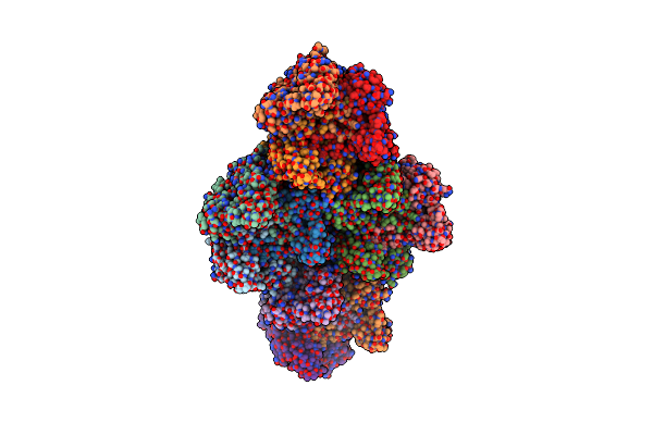 |
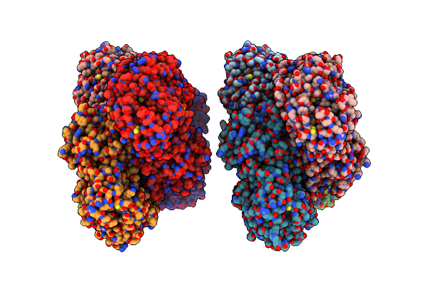
Quasi-Atomic Resolution Model Of Bacteriophage Prd1 Sus607 Mutant, Obtained By Combined Cryo-Em And X-Ray Crystallography.
Organism: Bacteriophage prd1
Method: ELECTRON MICROSCOPY Resolution:13.30 Å Release Date: 2002-03-15 Classification: VIRUS/VIRAL PROTEIN |
 |
Quasi-Atomic Resolution Model Of Bacteriophage Prd1 P3-Shell, Obtained By Combined Cryo-Em And X-Ray Crystallography.
Organism: Bacteriophage prd1
Method: ELECTRON MICROSCOPY Resolution:12.00 Å Release Date: 2001-12-05 Classification: VIRUS |
 |
Quasi-Atomic Resolution Model Of Bacteriophage Prd1 Sus1 Mutant, Obtained By Combined Cryo-Em And X-Ray Crystallography.
Organism: Bacteriophage prd1
Method: ELECTRON MICROSCOPY Resolution:14.00 Å Release Date: 2001-12-05 Classification: VIRUS |
 |
Quasi-Atomic Resolution Model Of Bacteriophage Prd1 Wild Type Virion, Obtained By Combined Cryo-Em And X-Ray Crystallography.
Organism: Bacteriophage prd1
Method: ELECTRON MICROSCOPY Resolution:25.00 Å Release Date: 2001-12-05 Classification: VIRUS |
 |
Organism: Enterobacteria phage prd1
Method: X-RAY DIFFRACTION Resolution:1.65 Å Release Date: 2001-01-24 Classification: VIRAL PROTEIN Ligands: CL, NA, MPD |