
Search Count: 12,742
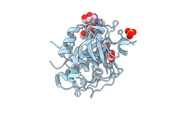 |
Organism: Ixodes ricinus
Method: X-RAY DIFFRACTION Release Date: 2025-12-17 Classification: HYDROLASE Ligands: SO4, E64 |
 |
Organism: Influenza a virus
Method: ELECTRON MICROSCOPY Release Date: 2025-12-17 Classification: VIRAL PROTEIN Ligands: A1IVV, NAG, CA |
 |
Organism: Influenza a virus
Method: ELECTRON MICROSCOPY Release Date: 2025-12-17 Classification: VIRAL PROTEIN Ligands: A1IVW, NAG, CA |
 |
Organism: Influenza a virus
Method: ELECTRON MICROSCOPY Release Date: 2025-12-17 Classification: VIRAL PROTEIN Ligands: A1IVX, NAG, CA |
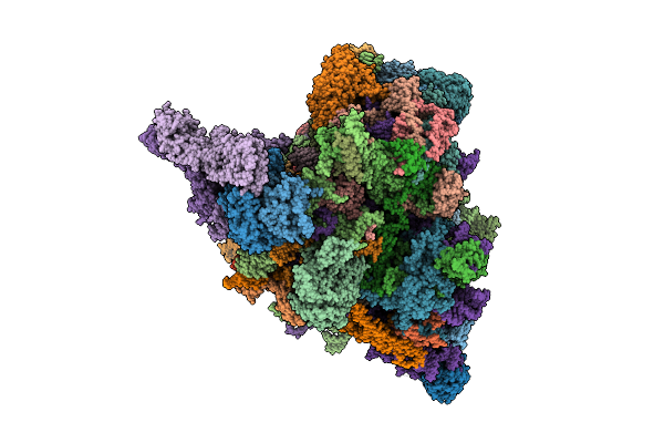 |
Translational Activators Aep1, Aep2 And Atp25 In Complex With Mrna And The Yeast Mitochondrial Ribosome
Organism: Saccharomyces cerevisiae w303
Method: ELECTRON MICROSCOPY Release Date: 2025-12-17 Classification: RIBOSOME Ligands: MG, GTP |
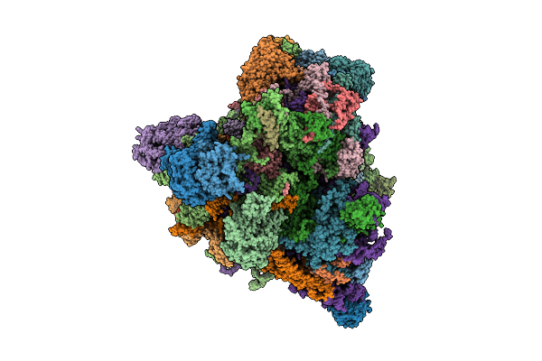 |
Translational Activator Aep3 In Complex With Mrna And The Yeast Mitochondrial Ribosome
Organism: Saccharomyces cerevisiae w303, Saccharomyces cerevisiae
Method: ELECTRON MICROSCOPY Release Date: 2025-12-17 Classification: RIBOSOME Ligands: MG, GTP |
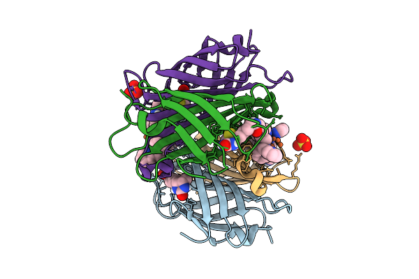 |
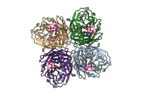
Streptavidin-E101Q-K121A-L124E Bound To Fe(Iii)-Biotin-Pentyl-Dipicolylamine Cofactor
Organism: Streptomyces avidinii
Method: X-RAY DIFFRACTION Release Date: 2025-12-17 Classification: METAL BINDING PROTEIN Ligands: A1BLH, FE, SO4 |
 |
Organism: Homo sapiens
Method: X-RAY DIFFRACTION Release Date: 2025-12-17 Classification: PROTEIN BINDING |
 |
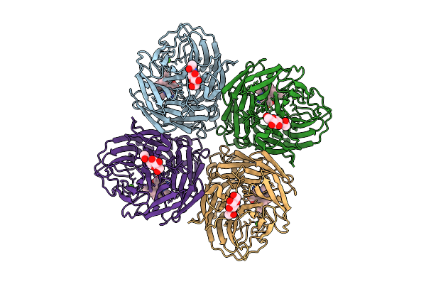
Organism: Vibrio cholerae
Method: ELECTRON MICROSCOPY Release Date: 2025-12-17 Classification: DNA BINDING PROTEIN |
 |
Organism: Vibrio cholerae
Method: ELECTRON MICROSCOPY Release Date: 2025-12-17 Classification: DNA BINDING PROTEIN |
 |
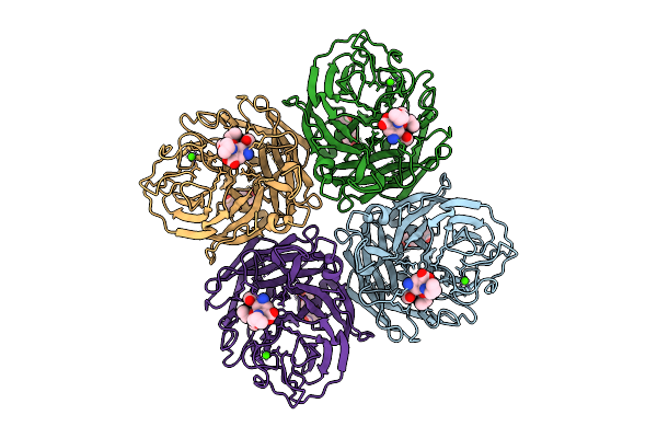
Organism: Vibrio cholerae
Method: ELECTRON MICROSCOPY Release Date: 2025-12-17 Classification: DNA BINDING PROTEIN |
 |
Organism: Vibrio cholerae
Method: ELECTRON MICROSCOPY Release Date: 2025-12-17 Classification: DNA BINDING PROTEIN/DNA |
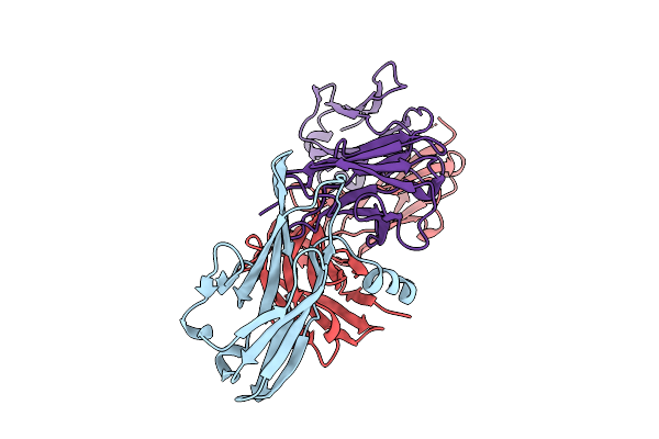 |
Crystal Structure Of Pilra In Complex With Fab Portion Of Antagonist Antibody
Organism: Homo sapiens
Method: X-RAY DIFFRACTION Release Date: 2025-12-17 Classification: SIGNALING PROTEIN |
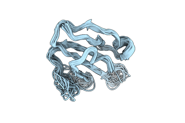 |
Solution Structure Of The Human Prostate Stem Cell Antigen (Psca), Water-Soluble Domain
|
 |
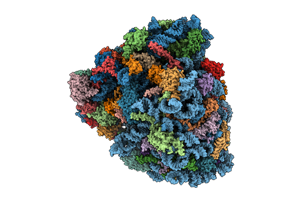
Organism: Homo sapiens, Oryctolagus cuniculus, Saccharomyces cerevisiae
Method: ELECTRON MICROSCOPY Release Date: 2025-12-17 Classification: RIBOSOME Ligands: SPD, ZN, GTP, PHE, ATP, MET, GCP, MG, K |
 |
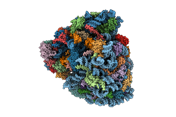
Organism: Homo sapiens, Oryctolagus cuniculus, Saccharomyces cerevisiae
Method: ELECTRON MICROSCOPY Release Date: 2025-12-17 Classification: RIBOSOME Ligands: SPD, ZN, GTP, PHE, ATP, MET, K, GCP, MG |
 |
Organism: Homo sapiens, Oryctolagus cuniculus, Saccharomyces cerevisiae
Method: ELECTRON MICROSCOPY Release Date: 2025-12-17 Classification: RIBOSOME Ligands: SPD, ZN, GTP, PHE, ATP, MET, GCP, MG, K |
 |
Organism: Homo sapiens, Oryctolagus cuniculus, Saccharomyces cerevisiae
Method: ELECTRON MICROSCOPY Release Date: 2025-12-17 Classification: RIBOSOME Ligands: SPD, ZN, GTP, PHE, ATP, MET, K, GCP, MG |
 |
Organism: Homo sapiens, Oryctolagus cuniculus
Method: ELECTRON MICROSCOPY Release Date: 2025-12-17 Classification: RIBOSOME Ligands: SPD, ZN, ATP, MET |
 |
Organism: Oryctolagus cuniculus, Homo sapiens
Method: ELECTRON MICROSCOPY Release Date: 2025-12-17 Classification: RIBOSOME Ligands: SPD, ZN, ATP, MET |