
Search Count: 48,323
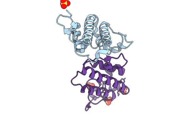 |
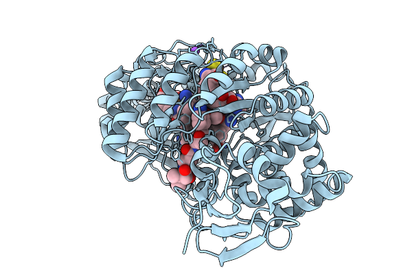
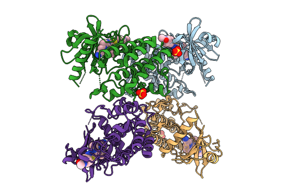
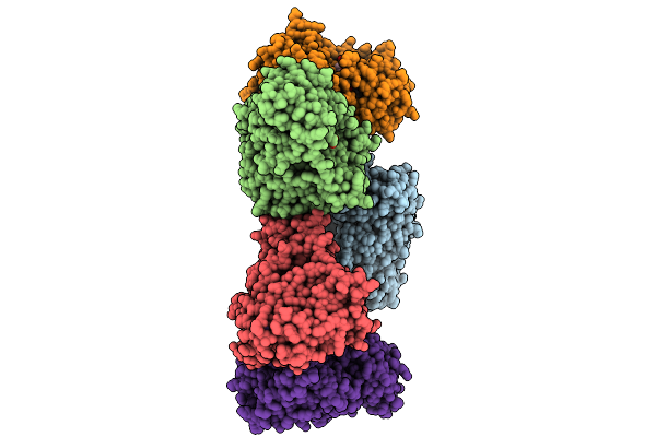
Crystal Structure Of Heterodimeric Crotoxin B From Crotalus Durissus Collilineatus
Organism: Crotalus durissus collilineatus
Method: X-RAY DIFFRACTION Resolution:1.89 Å Release Date: 2026-01-28 Classification: HYDROLASE Ligands: SO4 |
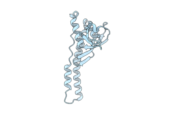 |
Structure Of A Chimeric Protein Composed Of The Snx5 Px Domain And The N-Terminal Region Of Ncoa7-As, Crystal Form I
Organism: Homo sapiens
Method: X-RAY DIFFRACTION Resolution:2.30 Å Release Date: 2026-01-28 Classification: UNKNOWN FUNCTION |
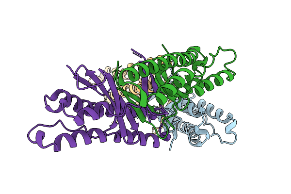 |
Structure Of A Chimeric Protein Composed Of The Snx5 Phox Domain And The N-Terminal Domain Of Ncoa7-As,Crystal Form Ii
Organism: Homo sapiens
Method: X-RAY DIFFRACTION Resolution:2.30 Å Release Date: 2026-01-28 Classification: UNKNOWN FUNCTION |
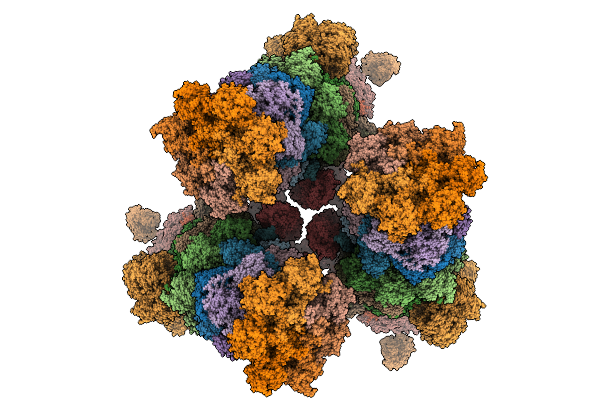 |
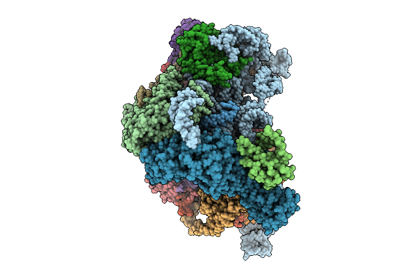
Organism: Saccharomyces cerevisiae s288c
Method: ELECTRON MICROSCOPY Release Date: 2026-01-28 Classification: HYDROLASE |
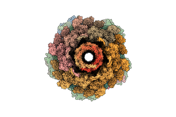 |
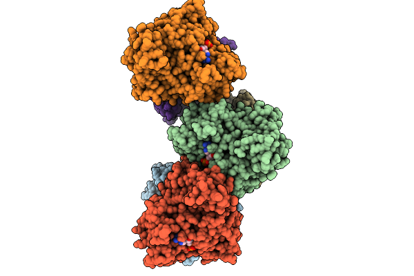
Organism: Escherichia phage t4
Method: ELECTRON MICROSCOPY Release Date: 2026-01-28 Classification: VIRAL PROTEIN |
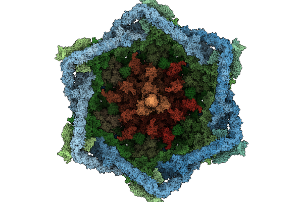 |
Organism: Escherichia phage t4
Method: ELECTRON MICROSCOPY Release Date: 2026-01-28 Classification: VIRAL PROTEIN Ligands: ZN |
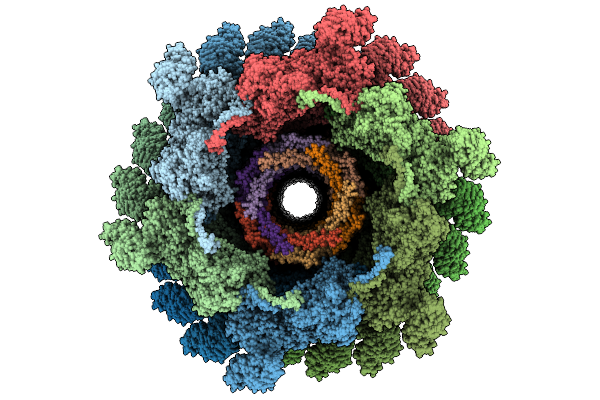 |
Organism: Escherichia phage t4
Method: ELECTRON MICROSCOPY Release Date: 2026-01-28 Classification: VIRAL PROTEIN |
 |
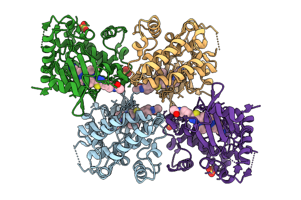
Crystal Structure Of Cyss From Corallococcus Sp. Ca054B With 5'-Deoxyadenosine, Methionine, And Pantetheinylated 3-Methoxyl-4-Amino Benzoic Acid Substrate Bound
Organism: Corallococcus sp. ca054b
Method: X-RAY DIFFRACTION Resolution:1.75 Å Release Date: 2026-01-28 Classification: OXIDOREDUCTASE/SUBSTRATE Ligands: B12, SF4, 5AD, MET, A1BUN, MG |
 |
Crystal Structure Of Cyss From Corallococcus Sp. Ca054B With 5'-Deoxyadenosine, Methionine, And Pantetheinylated 3-Ethoxy-4-Amino Benzoic Acid Substrate Bound
Organism: Corallococcus sp. ca054b
Method: X-RAY DIFFRACTION Resolution:2.00 Å Release Date: 2026-01-28 Classification: OXIDOREDUCTASE/SUBSTRATE Ligands: B12, SF4, 5AD, MET, A1BUP, NA |
 |
Crystal Structure Of Cyss From Corallococcus Sp. Ca054B With 5'-Deoxyadenosine And Methionine Bound
Organism: Corallococcus sp. ca054b
Method: X-RAY DIFFRACTION Resolution:1.94 Å Release Date: 2026-01-28 Classification: OXIDOREDUCTASE Ligands: B12, SF4, 5AD, MET, NA |
 |
Organism: Homo sapiens
Method: X-RAY DIFFRACTION Resolution:1.93 Å Release Date: 2026-01-28 Classification: TRANSFERASE/TRANSFERASE INHIBITOR Ligands: A1CKP, SO4 |
 |
Organism: Homo sapiens
Method: X-RAY DIFFRACTION Resolution:2.12 Å Release Date: 2026-01-28 Classification: TRANSFERASE/TRANSFERASE INHIBITOR Ligands: A1CKQ, SO4 |
 |
Organism: Homo sapiens
Method: X-RAY DIFFRACTION Resolution:2.43 Å Release Date: 2026-01-28 Classification: TRANSFERASE/TRANSFERASE INHIBITOR Ligands: A1CKS, SO4 |
 |
Crystal Structure Of The Human Retinoid X Receptor Dna-Binding Domain Bound To Trim16 Ir1 Response Element
Organism: Homo sapiens
Method: X-RAY DIFFRACTION Resolution:3.50 Å Release Date: 2026-01-28 Classification: TRANSCRIPTION Ligands: ZN |
 |
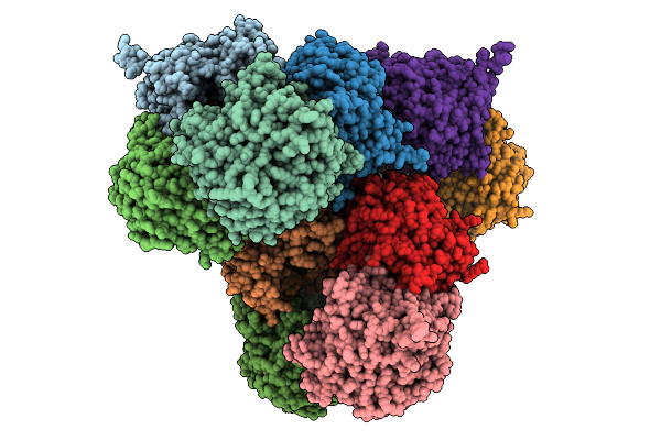
Organism: Saccharomyces cerevisiae
Method: ELECTRON MICROSCOPY Release Date: 2026-01-28 Classification: RIBOSOME |
 |
Organism: Prevotella
Method: X-RAY DIFFRACTION Resolution:2.50 Å Release Date: 2026-01-28 Classification: HYDROLASE Ligands: GOL |
 |
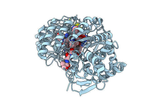
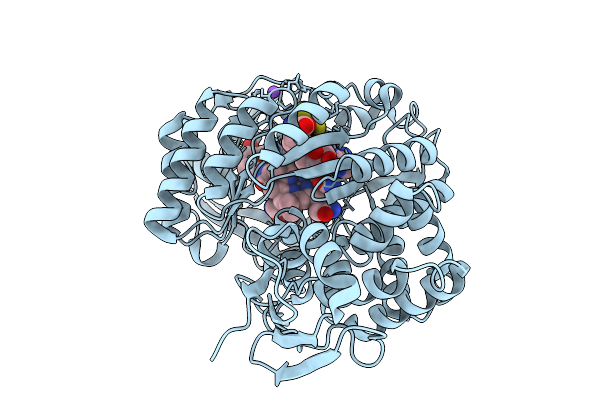
Sfx Crystal Structure Of Hen Egg-White Lysozyme (Hewl) Using A Guanosine Derivative Hydrogel As Crystal Matrix
Organism: Gallus gallus
Method: X-RAY DIFFRACTION Resolution:1.75 Å Release Date: 2026-01-28 Classification: HYDROLASE |
 |
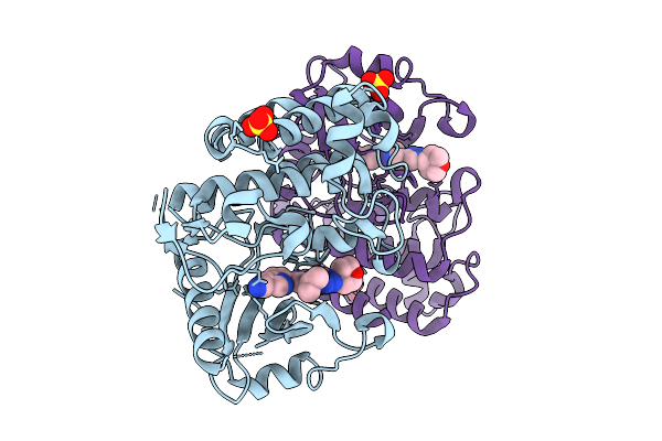
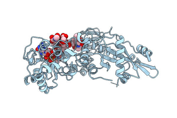
Crystal Structure Of Gamma-Glutamyl-Methylamide Synthetase From Methylovorus Mays (Mmgmas) In Complex With Atpgs
Organism: Methylovorus mays
Method: X-RAY DIFFRACTION Resolution:2.65 Å Release Date: 2026-01-28 Classification: LIGASE Ligands: AGS, MG |
 |
Crystal Structure Of Glutathione Transferase Iota 1 From Synechocystis Sp. Pcc 6803 In Complex With Fmn
Organism: Synechocystis sp. pcc 6803
Method: X-RAY DIFFRACTION Resolution:2.71 Å Release Date: 2026-01-28 Classification: TRANSFERASE Ligands: FMN, CL, NA |
 |
Organism: Arabidopsis thaliana
Method: X-RAY DIFFRACTION Resolution:1.41 Å Release Date: 2026-01-28 Classification: TRANSFERASE Ligands: GDP |