
Search Count: 6,285
 |
Organism: Rhodopseudomonas palustris
Method: X-RAY DIFFRACTION Release Date: 2025-10-29 Classification: FLUORESCENT PROTEIN Ligands: LBV |
 |
Organism: Streptomyces avidinii, Synthetic construct
Method: X-RAY DIFFRACTION Release Date: 2025-10-29 Classification: PROTEIN BINDING Ligands: GOL |
 |
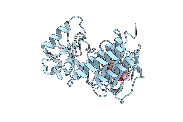
Human Pu.1 Ets-Domain (165-270) Bound To D(5'-Aataagcggaagtggg-3') D(5'-Tcccact*Cpd*Cgcttat-3')
Organism: Homo sapiens, Synthetic construct
Method: X-RAY DIFFRACTION Release Date: 2025-10-22 Classification: TRANSCRIPTION |
 |
Organism: Methylorubrum extorquens
Method: X-RAY DIFFRACTION Release Date: 2025-10-15 Classification: TRANSPORT PROTEIN Ligands: PQQ, CL |
 |
Organism: Candida albicans
Method: X-RAY DIFFRACTION Release Date: 2025-10-08 Classification: TRANSFERASE/INHIBITOR Ligands: PY1, CL |
 |
Organism: Candida albicans
Method: X-RAY DIFFRACTION Release Date: 2025-10-08 Classification: TRANSFERASE/INHIBITOR Ligands: CL, A1B7R, SO4 |
 |
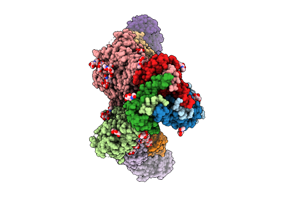
Organism: Human immunodeficiency virus 1
Method: ELECTRON MICROSCOPY Release Date: 2025-10-01 Classification: VIRAL PROTEIN Ligands: NAG |
 |
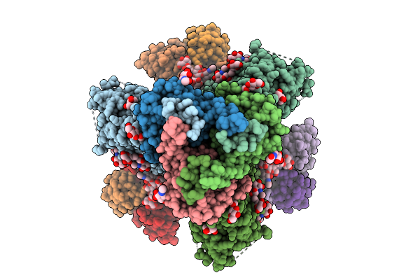
Organism: Human immunodeficiency virus 1, Homo sapiens
Method: ELECTRON MICROSCOPY Release Date: 2025-10-01 Classification: VIRAL PROTEIN/IMMUNE SYSTEM Ligands: NAG |
 |
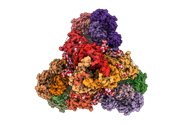
Organism: Human immunodeficiency virus 1, Homo sapiens
Method: ELECTRON MICROSCOPY Release Date: 2025-10-01 Classification: VIRAL PROTEIN/IMMUNE SYSTEM Ligands: NAG |
 |
Organism: Human immunodeficiency virus 1, Homo sapiens
Method: ELECTRON MICROSCOPY Release Date: 2025-10-01 Classification: VIRAL PROTEIN/IMMUNE SYSTEM Ligands: NAG |
 |
Organism: Human immunodeficiency virus 1, Homo sapiens
Method: ELECTRON MICROSCOPY Release Date: 2025-10-01 Classification: VIRAL PROTEIN/IMMUNE SYSTEM Ligands: NAG |
 |
Organism: Salpingoeca macrocollata
Method: X-RAY DIFFRACTION Release Date: 2025-10-01 Classification: IMMUNE SYSTEM Ligands: 1SY |
 |
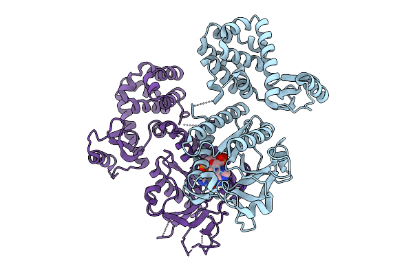
Organism: Pseudomonas aeruginosa
Method: X-RAY DIFFRACTION Release Date: 2025-10-01 Classification: ANTIVIRAL PROTEIN Ligands: ZN |
 |
Organism: Pseudomonas aeruginosa
Method: X-RAY DIFFRACTION Release Date: 2025-10-01 Classification: ANTIVIRAL PROTEIN Ligands: ZN, ATP, MG |
 |
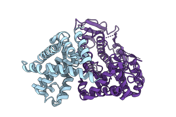
Structure Of Saro_1862, A Upf0261 Domain Protein From Novosphingobium Aromaticivorans With Bound Acetovanillone
Organism: Novosphingobium aromaticivorans dsm 12444
Method: X-RAY DIFFRACTION Release Date: 2025-09-24 Classification: HYDROLASE Ligands: I75, CIT, NA |
 |
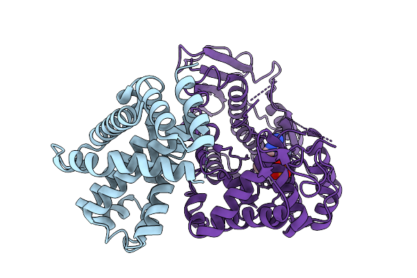
Organism: Homo sapiens
Method: X-RAY DIFFRACTION Release Date: 2025-09-24 Classification: HYDROLASE Ligands: MG, EDO, A1JD4 |
 |
Organism: Homo sapiens
Method: X-RAY DIFFRACTION Release Date: 2025-09-24 Classification: HYDROLASE Ligands: A1JED, TEW, MG |
 |
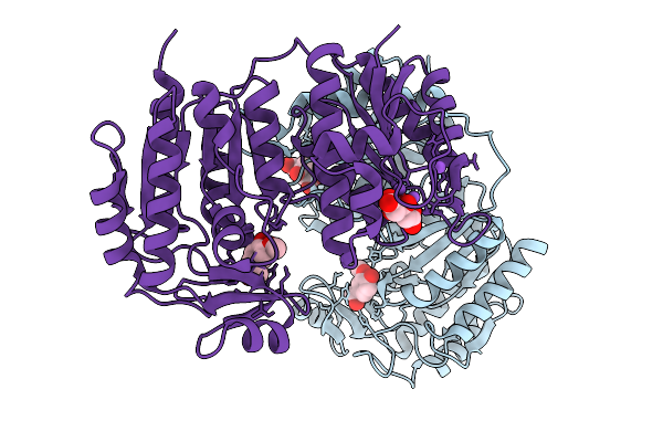
Tetrahydroprotoberberine N-Methyltransferase Complex With N-Methylcanadine And Sah
Organism: Glaucium flavum
Method: X-RAY DIFFRACTION Release Date: 2025-09-17 Classification: TRANSFERASE Ligands: SAH, A1A4I, SO4 |
 |
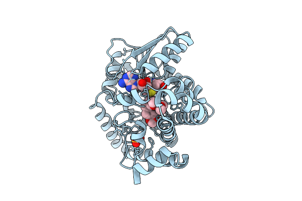
Tetrahydroprotoberberine N-Methyltransferase E204A Mutant In Complex With R-Reticuline And Sam
Organism: Glaucium flavum
Method: X-RAY DIFFRACTION Release Date: 2025-09-17 Classification: TRANSFERASE Ligands: SAM, A1A4H |
 |
Tetrahydroprotoberberine N-Methyltransferase E204A Mutant In Complex With (S)-Reticuline And Sam
Organism: Glaucium flavum
Method: X-RAY DIFFRACTION Release Date: 2025-09-17 Classification: TRANSFERASE Ligands: SAM, REN, GOL |