
Search Count: 30,425
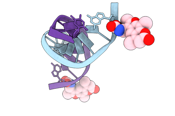 |
Photo-Crosslinkable Oligonucleotodes Which Have A Trioxsalen Conjugated To C2' Position Of Guanosine
Organism: Synthetic construct
Method: X-RAY DIFFRACTION Resolution:3.11 Å Release Date: 2026-01-28 Classification: DNA Ligands: A1L7U |
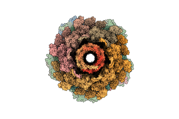 |
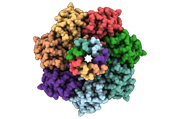
Organism: Escherichia phage t4
Method: ELECTRON MICROSCOPY Release Date: 2026-01-28 Classification: VIRAL PROTEIN |
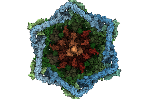 |
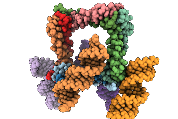
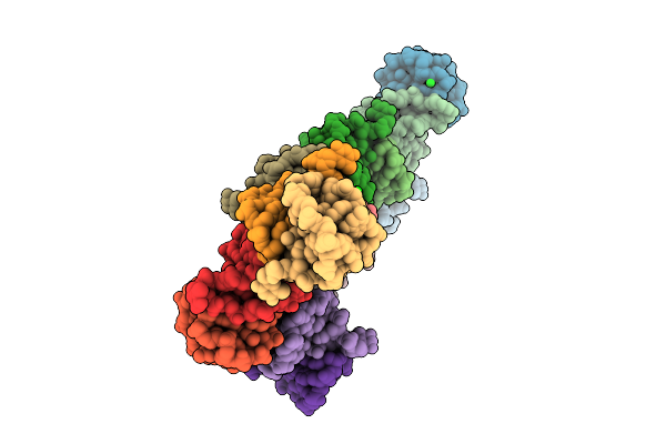
Organism: Escherichia phage t4
Method: ELECTRON MICROSCOPY Release Date: 2026-01-28 Classification: VIRAL PROTEIN Ligands: ZN |
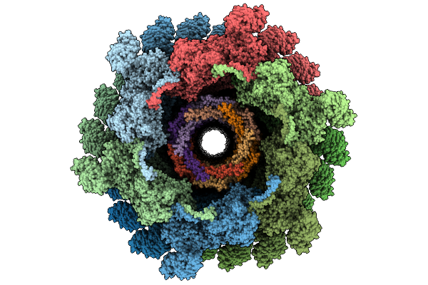 |
Organism: Escherichia phage t4
Method: ELECTRON MICROSCOPY Release Date: 2026-01-28 Classification: VIRAL PROTEIN |
 |
Organism: Escherichia coli
Method: ELECTRON MICROSCOPY Resolution:2.99 Å Release Date: 2026-01-28 Classification: MEMBRANE PROTEIN Ligands: D21 |
 |
Organism: Hepatitis delta virus, Synthetic construct
Method: ELECTRON MICROSCOPY Resolution:3.50 Å Release Date: 2026-01-28 Classification: VIRUS |
 |
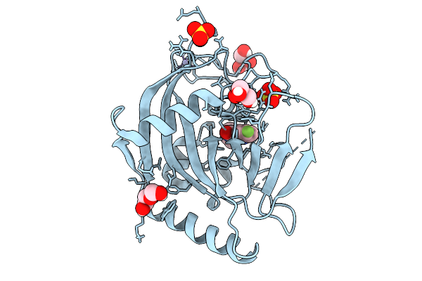
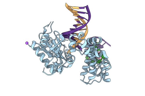
Organism: Homo sapiens
Method: X-RAY DIFFRACTION Resolution:1.89 Å Release Date: 2026-01-28 Classification: TRANSCRIPTION Ligands: A1JIF, PEG, ZN, SO4 |
 |
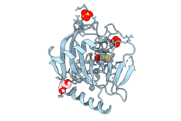
Organism: Homo sapiens
Method: X-RAY DIFFRACTION Resolution:2.05 Å Release Date: 2026-01-28 Classification: TRANSFERASE Ligands: GOL, PEG, A1JI1, ZN, SO4 |
 |
Sam-Dependent C6-Fpp Methytransferase From Streptomyces Varsoviensis In Complex With Sah And Fpp
Organism: Streptomyces varsoviensis
Method: X-RAY DIFFRACTION Resolution:2.20 Å Release Date: 2026-01-28 Classification: TRANSFERASE Ligands: FPP, SAH, PG4, PGE, PE8, PEG, EDO |
 |
Organism: Homo sapiens
Method: X-RAY DIFFRACTION Resolution:1.90 Å Release Date: 2026-01-28 Classification: TRANSFERASE Ligands: DQV, NCA |
 |
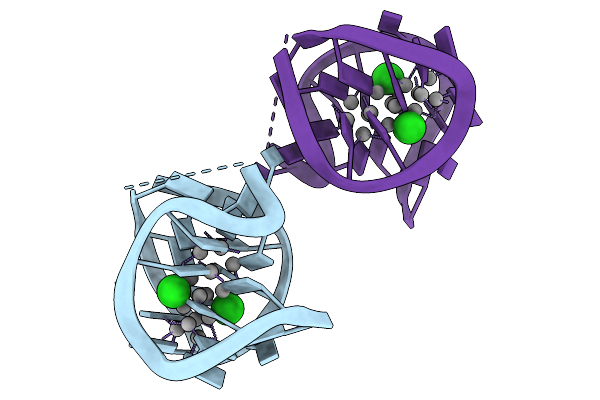
Probing Positions 3 And 8: Insights Into A 960 Nm Emissive Dna-Stabilized Silver Nanocluster
Organism: Synthetic construct
Method: X-RAY DIFFRACTION Resolution:1.90 Å Release Date: 2026-01-28 Classification: DNA Ligands: AG, CL, SR |
 |
Crystal Structure Of Single-Strand Dna-Stabilized Ag16 Nanocluster: T5 Linker
Organism: Synthetic construct
Method: X-RAY DIFFRACTION Resolution:2.00 Å Release Date: 2026-01-28 Classification: DNA Ligands: AG, CL |
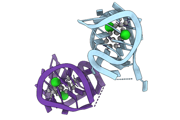 |
Crystal Structure Of Single-Strand Dna-Stabilized Ag16 Nanocluster: T6 Linker
Organism: Synthetic construct
Method: X-RAY DIFFRACTION Resolution:1.70 Å Release Date: 2026-01-28 Classification: DNA Ligands: CL, AG |
 |
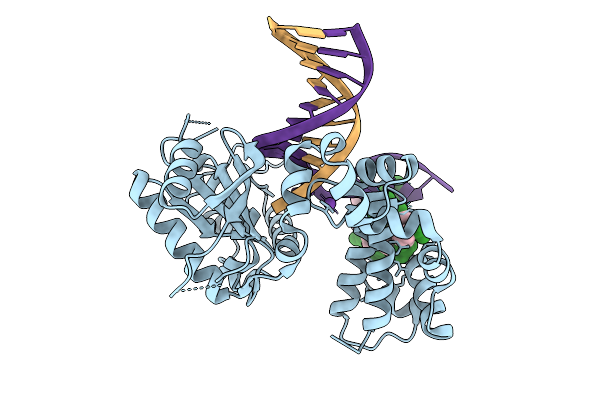
Organism: Homo sapiens, Synthetic construct
Method: X-RAY DIFFRACTION Resolution:1.85 Å Release Date: 2026-01-28 Classification: REPLICATION, HYDROLASE/DNA |
 |
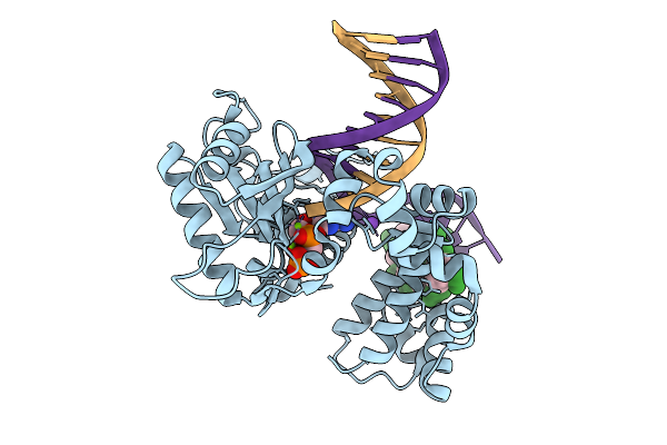
Organism: Homo sapiens, Synthetic construct
Method: X-RAY DIFFRACTION Resolution:1.80 Å Release Date: 2026-01-28 Classification: REPLICATION, HYDROLASE/DNA |
 |
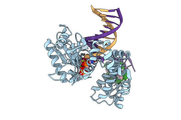
Organism: Homo sapiens, Synthetic construct
Method: X-RAY DIFFRACTION Resolution:1.85 Å Release Date: 2026-01-28 Classification: REPLICATION, HYDROLASE/DNA |
 |
Organism: Homo sapiens, Synthetic construct
Method: X-RAY DIFFRACTION Resolution:1.80 Å Release Date: 2026-01-28 Classification: REPLICATION, HYDROLASE/DNA |
 |
Organism: Homo sapiens, Synthetic construct
Method: X-RAY DIFFRACTION Resolution:1.90 Å Release Date: 2026-01-28 Classification: REPLICATION, HYDROLASE/DNA |
 |
Organism: Homo sapiens, Synthetic construct
Method: X-RAY DIFFRACTION Resolution:1.93 Å Release Date: 2026-01-28 Classification: REPLICATION, HYDROLASE/DNA |
 |
Organism: Homo sapiens, Synthetic construct
Method: X-RAY DIFFRACTION Resolution:1.90 Å Release Date: 2026-01-28 Classification: REPLICATION, HYDROLASE/DNA |