
Search Count: 77,205
 |
Organism: Homo sapiens
Method: ELECTRON MICROSCOPY Release Date: 2025-12-31 Classification: HYDROLASE Ligands: ATP, ADP |
 |
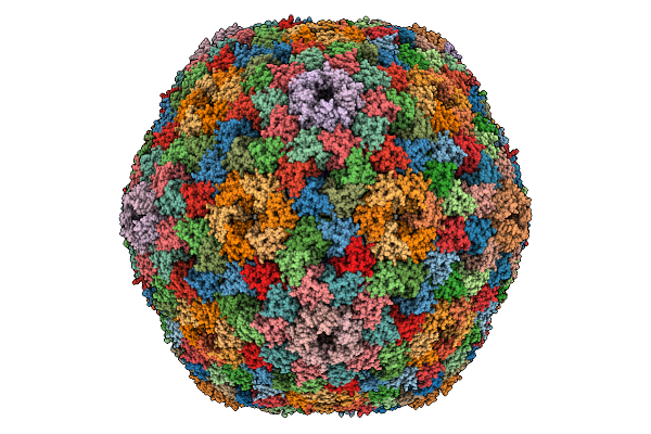
Organism: Porcine epidemic diarrhea virus
Method: X-RAY DIFFRACTION Release Date: 2025-12-31 Classification: VIRAL PROTEIN |
 |
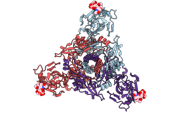
Hexamer Msp1 From S.Cerevisiae(With A Catalytic Dead Mutation) In Complex With An Unknown Peptide Substrate
Organism: Saccharomyces cerevisiae (strain atcc 204508 / s288c), Escherichia coli bl21(de3)
Method: ELECTRON MICROSCOPY Release Date: 2025-12-31 Classification: MEMBRANE PROTEIN Ligands: ATP, MG |
 |
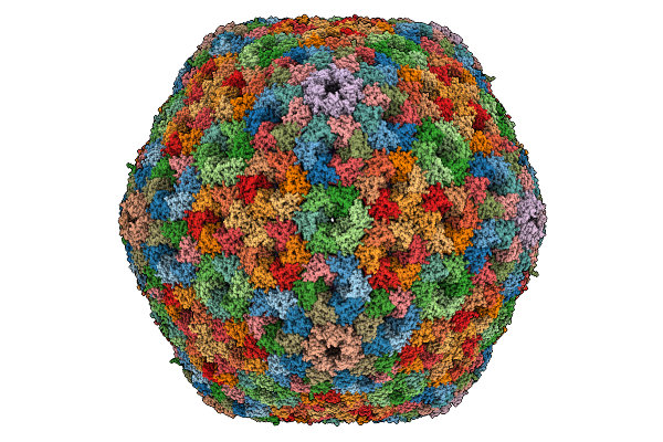
Organism: Porcine epidemic diarrhea virus
Method: X-RAY DIFFRACTION Release Date: 2025-12-31 Classification: VIRAL PROTEIN |
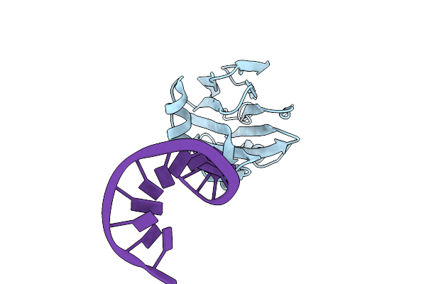 |
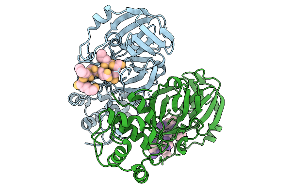
Structure Of Gh-Tdh With Additional N-Terminus In Complex With Double-Stranded Dna Containing A 2-Nucleotide 5' Overhang
Organism: Grimontia hollisae, Synthetic construct
Method: X-RAY DIFFRACTION Release Date: 2025-12-31 Classification: TOXIN |
 |
Organism: Homo sapiens
Method: ELECTRON MICROSCOPY Release Date: 2025-12-31 Classification: TRANSPORT PROTEIN Ligands: BG6 |
 |
Organism: Homo sapiens
Method: ELECTRON MICROSCOPY Release Date: 2025-12-31 Classification: TRANSPORT PROTEIN Ligands: A1EG7 |
 |
Organism: Homo sapiens
Method: ELECTRON MICROSCOPY Release Date: 2025-12-31 Classification: TRANSPORT PROTEIN Ligands: LMN |
 |
Organism: Homo sapiens
Method: ELECTRON MICROSCOPY Release Date: 2025-12-31 Classification: TRANSPORT PROTEIN Ligands: LMN, PO4 |
 |
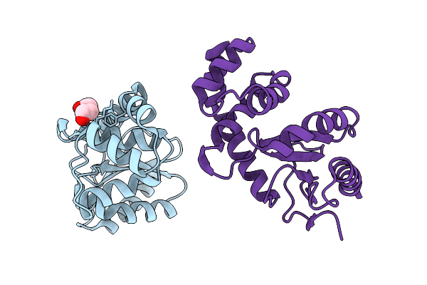
Cryo-Em Structure Of The Cytosolic Armh2-Efcab9-Catsperz Subcomplex Of The Mouse Catspermasome
Organism: Mus musculus
Method: ELECTRON MICROSCOPY Release Date: 2025-12-31 Classification: CYTOSOLIC PROTEIN |
 |
Organism: Halothiobacillus neapolitanus c2
Method: ELECTRON MICROSCOPY Release Date: 2025-12-31 Classification: STRUCTURAL PROTEIN |
 |
Cryo-Em Structure Of Carboxysomal Mid-Shell: T = 16 Shell Under C1 Symmetry.
Organism: Halothiobacillus neapolitanus c2
Method: ELECTRON MICROSCOPY Release Date: 2025-12-31 Classification: STRUCTURAL PROTEIN |
 |
The Psi3-Isia43 Complex With A Closed Double Ring Of Isia Proteins Bound To A Trimeric Psi Core
Organism: Thermosynechococcus vestitus bp-1
Method: ELECTRON MICROSCOPY Release Date: 2025-12-31 Classification: PHOTOSYNTHESIS Ligands: CLA, PQN, SF4, BCR, LHG, LMU, SQD, LMG, CA |
 |
The Psi1-Isia13 Complex With Double-Layered Isia Proteins Bound To The Monomeric Psi Core
Organism: Thermosynechococcus vestitus bp-1
Method: ELECTRON MICROSCOPY Release Date: 2025-12-31 Classification: PHOTOSYNTHESIS Ligands: CLA, BCR, SQD, LMG, LHG, PQN, SF4, LMU |
 |
Organism: Severe acute respiratory syndrome coronavirus 2
Method: ELECTRON MICROSCOPY Release Date: 2025-12-31 Classification: VIRAL PROTEIN Ligands: NAG |
 |
Organism: Human gammaherpesvirus 4
Method: ELECTRON MICROSCOPY Release Date: 2025-12-31 Classification: VIRAL PROTEIN |
 |
Crystal Structure Of Gitr In Complex With Ligand-Non-Competitive Ab#1 Fab Fragment
Organism: Homo sapiens
Method: X-RAY DIFFRACTION Release Date: 2025-12-31 Classification: IMMUNE SYSTEM Ligands: ACT, NAG |
 |
Organism: Bordetella pertussis
Method: X-RAY DIFFRACTION Release Date: 2025-12-31 Classification: OXIDOREDUCTASE Ligands: PEG |
 |
Organism: Sus scrofa
Method: ELECTRON MICROSCOPY Release Date: 2025-12-31 Classification: SIGNALING PROTEIN Ligands: E2Q |
 |
Organism: Homo sapiens
Method: ELECTRON MICROSCOPY Release Date: 2025-12-31 Classification: HYDROLASE Ligands: ATP, ADP |