
Search Count: 22,685
 |
Organism: Homo sapiens
Method: X-RAY DIFFRACTION Release Date: 2025-11-05 Classification: HYDROLASE/INHIBITOR |
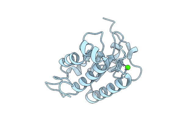 |
Organism: Chlamydomonas reinhardtii
Method: X-RAY DIFFRACTION Release Date: 2025-11-05 Classification: PHOTOSYNTHESIS Ligands: CA |
 |
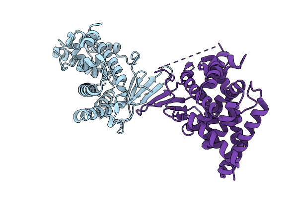
Organism: Lama glama, Homo sapiens
Method: X-RAY DIFFRACTION Release Date: 2025-11-05 Classification: GENE REGULATION |
 |
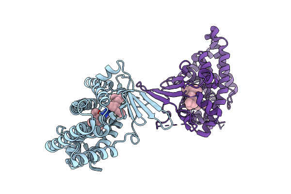
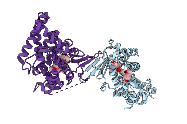
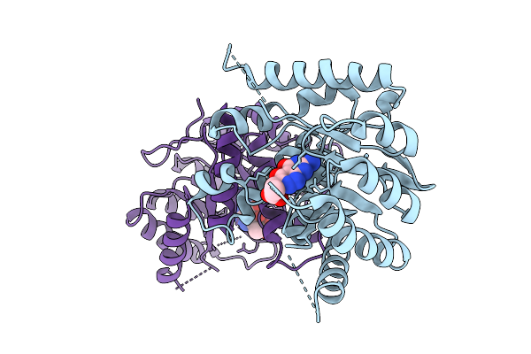
K115 Acetylated Human Muscle Pyruvate Kinase, Isoform M2 (Pkm2), In Complex With Fbp
Organism: Homo sapiens
Method: X-RAY DIFFRACTION Release Date: 2025-11-05 Classification: TRANSFERASE Ligands: FBP, EDO, SIN, GOL, K |
 |
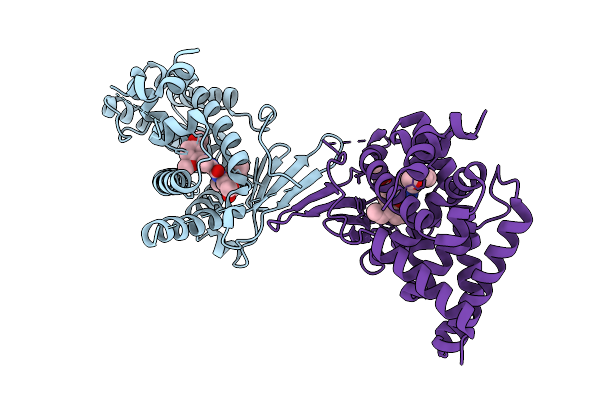
Organism: Homo sapiens
Method: X-RAY DIFFRACTION Release Date: 2025-11-05 Classification: TRANSFERASE Ligands: GOL, EDO, MG, TRS, SIN, K |
 |
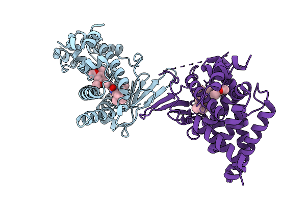
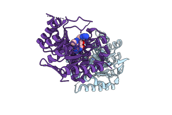
K166 Acetylated Human Muscle Pyruvate Kinase, Isoform M2 (Pkm2), In Complex With Fbp
Organism: Homo sapiens
Method: X-RAY DIFFRACTION Release Date: 2025-11-05 Classification: TRANSFERASE Ligands: FBP, MG |
 |
Crystal Structure Of The L411A Mutant Of Pregnane X Receptor Ligand Binding Domain (Apo Form)
Organism: Homo sapiens
Method: X-RAY DIFFRACTION Release Date: 2025-11-05 Classification: TRANSCRIPTION |
 |
Crystal Structure Of The L411A Mutant Of Pregnane X Receptor Ligand Binding Domain In Complex With Sjpyt-328
Organism: Homo sapiens
Method: X-RAY DIFFRACTION Release Date: 2025-11-05 Classification: TRANSCRIPTION Ligands: WU2 |
 |
Crystal Structure Of The L411A Mutant Of Pregnane X Receptor Ligand Binding Domain In Complex With Sjpyt-331
Organism: Homo sapiens
Method: X-RAY DIFFRACTION Release Date: 2025-11-05 Classification: TRANSCRIPTION Ligands: WU6 |
 |
Crystal Structure Of The L411W Mutant Of Pregnane X Receptor Ligand Binding Domain (Apo Form)
Organism: Homo sapiens
Method: X-RAY DIFFRACTION Release Date: 2025-11-05 Classification: TRANSCRIPTION |
 |
Crystal Structure Of The L411W Mutant Of Pregnane X Receptor Ligand Binding Domain In Complex With Sjpyt-328
Organism: Homo sapiens
Method: X-RAY DIFFRACTION Release Date: 2025-11-05 Classification: TRANSCRIPTION Ligands: WU2 |
 |
Crystal Structure Of The L411W Mutant Of Pregnane X Receptor Ligand Binding Domain In Complex With Sjpyt-331
Organism: Homo sapiens
Method: X-RAY DIFFRACTION Release Date: 2025-11-05 Classification: TRANSCRIPTION Ligands: WU6 |
 |
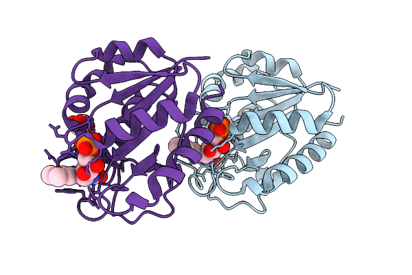
Organism: Escherichia coli
Method: X-RAY DIFFRACTION Release Date: 2025-11-05 Classification: TRANSFERASE Ligands: A1CG3 |
 |
Organism: Escherichia coli
Method: X-RAY DIFFRACTION Release Date: 2025-11-05 Classification: TRANSFERASE Ligands: A1CG7 |
 |
E20K/N28G/V36L/D43K/Q48E/I59A/E61K/E72K/V76L/N79S/I92A/D126K/A142V/D153K/D154E/S158T Flavodoxin From Anabaena
Organism: Nostoc sp. pcc 7119
Method: X-RAY DIFFRACTION Release Date: 2025-11-05 Classification: ELECTRON TRANSPORT Ligands: FMN, GOL |
 |
Spindlin Family Member 4 With Crystallization Epitope Mutations G129S:H131E
Organism: Homo sapiens
Method: X-RAY DIFFRACTION Release Date: 2025-11-05 Classification: PEPTIDE BINDING PROTEIN |
 |
Crystal Structure Of The Mycobacterium Tuberculosis Regulator Virs (N-Terminal Fragment 4-208) In Complex With The Drug Candidate Alpibectir
Organism: Mycobacterium tuberculosis h37rv
Method: X-RAY DIFFRACTION Release Date: 2025-10-29 Classification: TRANSCRIPTION Ligands: YPQ |
 |
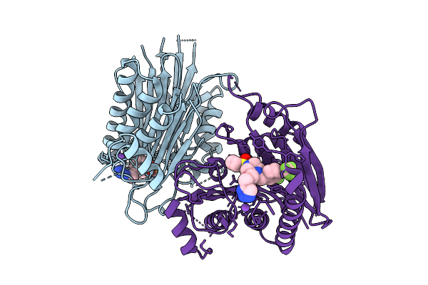
Glucagon Like Peptide Receptor-1 (Glp1R) A316T Mutant With Glp-1 Peptide. Dominant Negative Gs Complex.
Organism: Homo sapiens, Lama glama
Method: ELECTRON MICROSCOPY Release Date: 2025-10-29 Classification: MEMBRANE PROTEIN |
 |
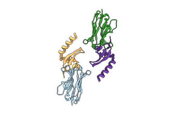
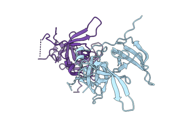
Organism: Homo sapiens, Camelidae
Method: X-RAY DIFFRACTION Release Date: 2025-10-29 Classification: IMMUNE SYSTEM Ligands: GOL |
 |
Organism: Homo sapiens, Camelidae
Method: X-RAY DIFFRACTION Release Date: 2025-10-29 Classification: IMMUNE SYSTEM |