
Search Count: 11,053
 |
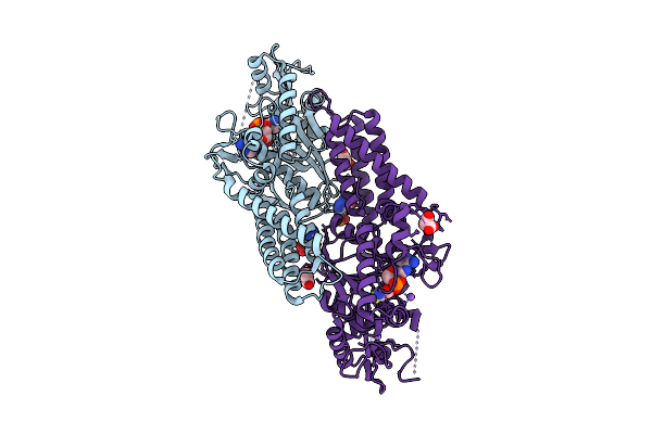
Organism: Escherichia coli, Synthetic construct
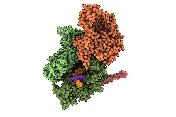
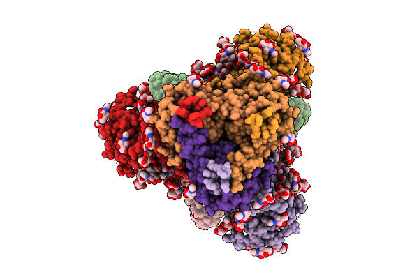
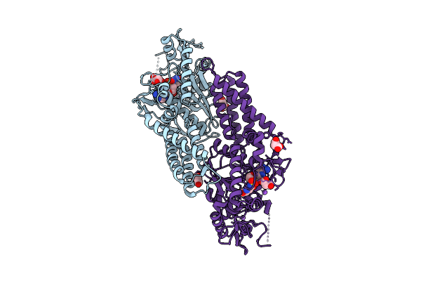
Method: ELECTRON MICROSCOPY Release Date: 2025-11-05 Classification: TRANSCRIPTION Ligands: MG, ZN |
 |
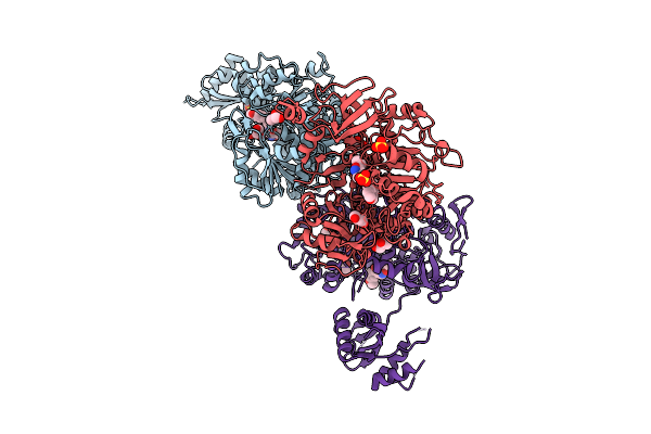
Organism: Escherichia coli, Synthetic construct
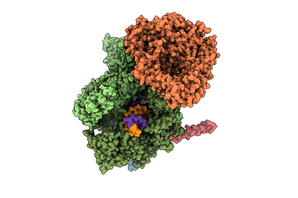
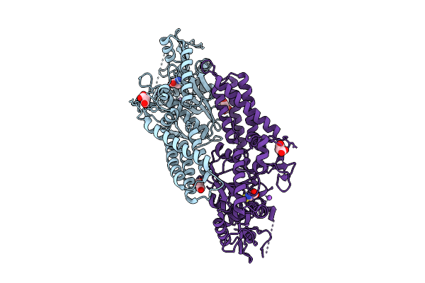
Method: ELECTRON MICROSCOPY Release Date: 2025-11-05 Classification: TRANSCRIPTION Ligands: MG, ZN |
 |
Organism: Escherichia coli, Synthetic construct
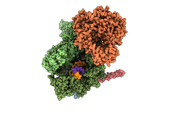
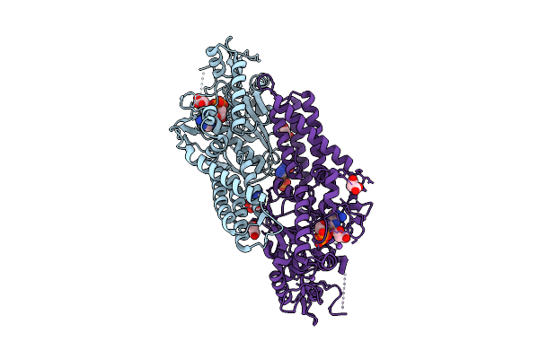
Method: ELECTRON MICROSCOPY Release Date: 2025-11-05 Classification: TRANSCRIPTION Ligands: MG, ZN |
 |
Organism: Escherichia coli, Synthetic construct
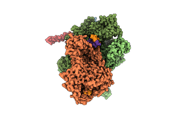
Method: ELECTRON MICROSCOPY Release Date: 2025-11-05 Classification: TRANSCRIPTION Ligands: MG, ZN |
 |
Organism: Escherichia coli, Synthetic construct
Method: ELECTRON MICROSCOPY Release Date: 2025-11-05 Classification: TRANSCRIPTION Ligands: MG, ZN |
 |
Organism: Escherichia coli, Synthetic construct
Method: ELECTRON MICROSCOPY Release Date: 2025-11-05 Classification: TRANSCRIPTION Ligands: MG, ZN |
 |
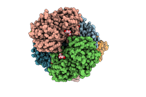
Organism: Mus musculus
Method: ELECTRON MICROSCOPY Release Date: 2025-11-05 Classification: RIBOSOME Ligands: ZN, MG |
 |
Organism: Yersinia ruckeri
Method: X-RAY DIFFRACTION Release Date: 2025-11-05 Classification: METAL BINDING PROTEIN Ligands: FE |
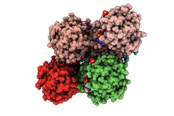 |
Organism: Yersinia ruckeri
Method: X-RAY DIFFRACTION Release Date: 2025-11-05 Classification: METAL BINDING PROTEIN Ligands: FE, A1I15, SO4 |
 |
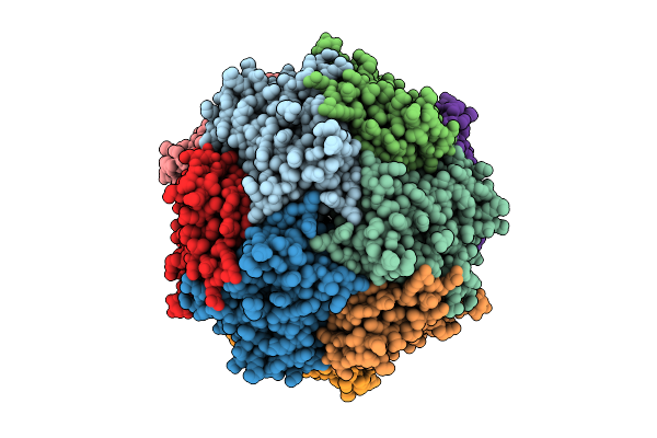
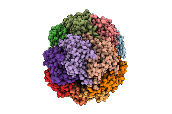
Organism: Escherichia phage ms2
Method: ELECTRON MICROSCOPY Release Date: 2025-11-05 Classification: VIRUS LIKE PARTICLE |
 |
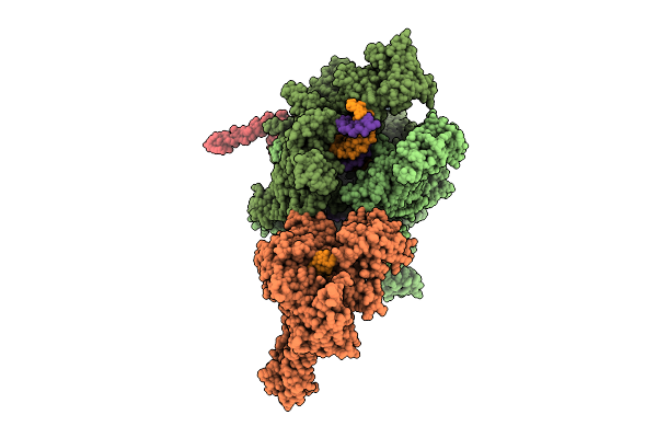
Cryo-Em Structure Of Vaccine-Elicited Antibody T3_Qb_G12 In Complex With Hiv Env Trimer Q23-Apex-Gt1
Organism: Mus musculus, Homo sapiens
Method: ELECTRON MICROSCOPY Release Date: 2025-11-05 Classification: VIRAL PROTEIN/IMMUNE SYSTEM Ligands: NAG |
 |
Crystal Structure Of Cysteinyl-Trna Synthetase (Cysrs) From Plasmodium Falciparum In Complex With O5'-(L-Glutamyl-Sulfamoyl)-Adenosine
Organism: Plasmodium falciparum 3d7
Method: X-RAY DIFFRACTION Release Date: 2025-11-05 Classification: TRANSFERASE Ligands: MLI, GSU, ZN, NA |
 |
Crystal Structure Of Cysteinyl-Trna Synthetase (Cysrs) From Plasmodium Falciparum In Complex With Cysteine
Organism: Plasmodium falciparum 3d7
Method: X-RAY DIFFRACTION Release Date: 2025-11-05 Classification: TRANSFERASE Ligands: MLI, CYS, ZN, NA |
 |
Crystal Structure Of Cysteinyl-Trna Synthetase (Cysrs) From Plasmodium Falciparum In Complex With Adp
Organism: Plasmodium falciparum 3d7
Method: X-RAY DIFFRACTION Release Date: 2025-11-05 Classification: TRANSFERASE Ligands: ADP, MLI, ZN, NA |
 |
Crystal Structure Of Cysteinyl-Trna Synthetase (Cysrs) From Plasmodium Falciparum In Complex With Amp And Cysteine
Organism: Plasmodium falciparum 3d7
Method: X-RAY DIFFRACTION Release Date: 2025-11-05 Classification: TRANSFERASE Ligands: CYS, AMP, MLI, ZN, NA |
 |
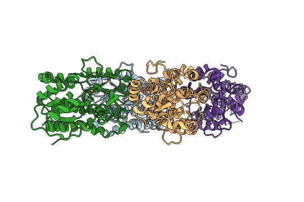
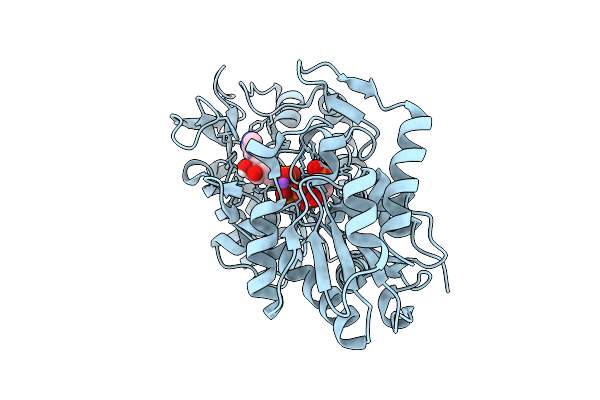
Crystal Structure Of A Glyceraldehyde-3-Phosphate Dehydrogenase From Bordetella Pertussis (Monoclinic P Form)
Organism: Bordetella pertussis tohama i
Method: X-RAY DIFFRACTION Release Date: 2025-11-05 Classification: OXIDOREDUCTASE Ligands: MG, PGE, PG4, PEG, GOL, TRS |
 |
Crystal Structure Of Acetyl-Coa Synthetase From Cryptococcus Neoformans H99 In Complex With Inhibitor Hgn-1310 (Dd3-027)
Organism: Cryptococcus neoformans var. grubii
Method: X-RAY DIFFRACTION Release Date: 2025-10-29 Classification: LIGASE Ligands: GOL, SO4, CL, A1AV1 |
 |
Structure Of Thioferritin (Pfdpsl) With Ferrihydrite Growth At A Single Three-Fold Pore.
Organism: Pyrococcus furiosus
Method: ELECTRON MICROSCOPY Release Date: 2025-10-29 Classification: METAL BINDING PROTEIN Ligands: FE, OXY, O |
 |
Human Phosphoglycerate Kinase In With Mixture Of Products And Substrates Produced By Cross-Soaking A Tsa Crystal
Organism: Homo sapiens
Method: X-RAY DIFFRACTION Release Date: 2025-10-29 Classification: TRANSFERASE Ligands: ATP, X15, NA, CL |
 |
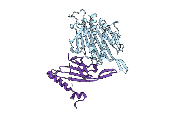
Crystal Structure Of S. Epidermidis Clpp In Complex With Tavaborole - Soaking
Organism: Staphylococcus epidermidis
Method: X-RAY DIFFRACTION Release Date: 2025-10-29 Classification: HYDROLASE Ligands: ACT, A1II0 |