
Search Count: 1,069
 |
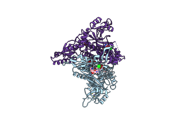
Organism: Mycobacterium tuberculosis h37rv
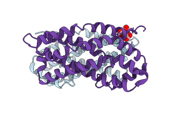
Method: X-RAY DIFFRACTION Release Date: 2025-10-08 Classification: TRANSPORT PROTEIN Ligands: ATP, MG |
 |
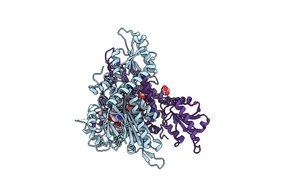
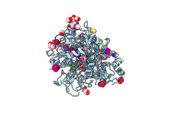
Crystal Structure Of Lysyl-Trna Synthetase From Plasmodium Falciparum Complexed With Lys-Ams
Organism: Plasmodium falciparum
Method: X-RAY DIFFRACTION Release Date: 2025-09-10 Classification: LIGASE Ligands: KAA |
 |
High Resolution Structure Of Lectin-Like Ox-Ldl Receptor 1 With Bi-0115 In Space Group P 21 21 21
Organism: Homo sapiens
Method: X-RAY DIFFRACTION Release Date: 2025-07-23 Classification: LIPID BINDING PROTEIN Ligands: NJT, GOL |
 |
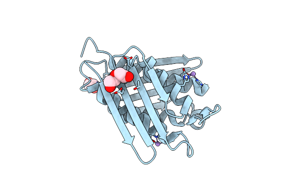
Structural Insight Into Dna Recognition By Rrm1+2 Domain Of Human Etr-3 Protein
Organism: Homo sapiens
Method: X-RAY DIFFRACTION Resolution:1.82 Å Release Date: 2025-05-28 Classification: RNA BINDING PROTEIN Ligands: MG |
 |
Organism: Plasmodium falciparum
Method: X-RAY DIFFRACTION Resolution:2.13 Å Release Date: 2025-05-21 Classification: STRUCTURAL PROTEIN |
 |
Crystal Structure Of Dh Domain Of Fyve Domain Containing Protein(Fp10) From Entamoeba Histolytica
Organism: Entamoeba histolytica hm-1:imss-a
Method: X-RAY DIFFRACTION Resolution:2.48 Å Release Date: 2025-05-21 Classification: SIGNALING PROTEIN Ligands: CIT |
 |
Crystal Structure Of The Complex Of Camel Peptidoglycan Recognition Protein, Pgrp-S With Malic Acid And Oxalic Acid At 2.3 A Resolution
Organism: Camelus dromedarius
Method: X-RAY DIFFRACTION Resolution:2.31 Å Release Date: 2025-05-14 Classification: IMMUNE SYSTEM Ligands: MLT, OXD |
 |
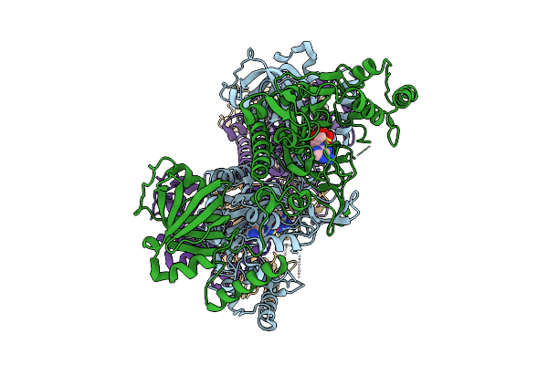
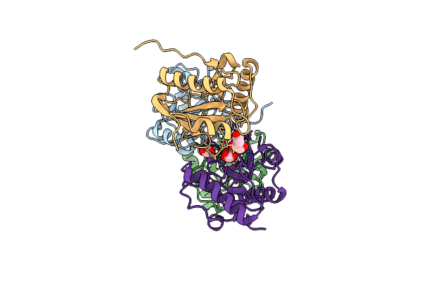
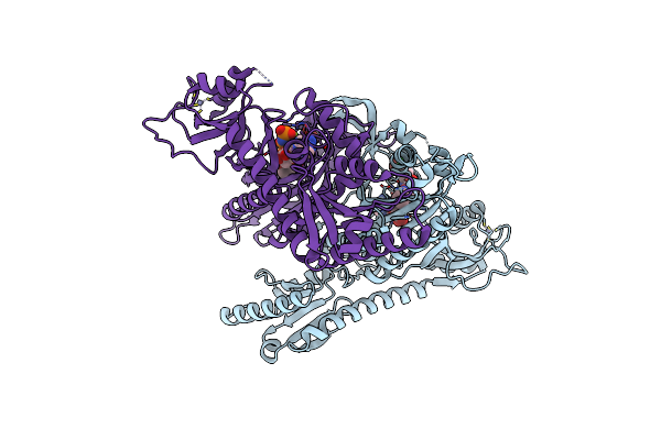
Cryo-Em Structure Of Pyruvate Carboxylase From Mycobacterium Tuberculosis In Complex With Acetyl-Coa And Adp
Organism: Mycobacterium tuberculosis h37rv
Method: ELECTRON MICROSCOPY Release Date: 2025-05-07 Classification: LIGASE Ligands: ADP, ZN, ACO |
 |
Structure Of The Intermediate Of Lactoperoxidase Formed With Thiocynate And Hydrogen Peroxidase At 1.99 A Resolution.
Organism: Bos taurus
Method: X-RAY DIFFRACTION Resolution:2.00 Å Release Date: 2025-03-26 Classification: OXIDOREDUCTASE Ligands: HEM, CA, NAG, IOD, SCN, PEO, GOL |
 |
Crystal Structure Of The Complex Of Lactoperoxidase With Nitric Oxide At 1.72 A Resolution
Organism: Bos taurus
Method: X-RAY DIFFRACTION Resolution:1.72 Å Release Date: 2024-12-18 Classification: OXIDOREDUCTASE Ligands: NAG, IOD, SCN, PGE, NO2, NO, HEM, CA |
 |
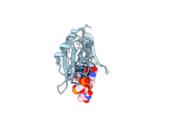
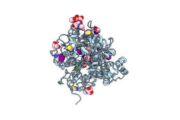
Structure Of Phosphopantetheine Adenylyltransferase (Ppat) From Enterobacter Spp. With The Expression Tag Bound In The Substrate Binding Site Of A Neighbouring Molecule At 2.20 A Resolution.
Organism: Enterobacter sp. 638
Method: X-RAY DIFFRACTION Release Date: 2024-12-11 Classification: TRANSFERASE Ligands: PAE, GOL |
 |
Crystal Structure Of Anopheles Culicifacies Prolyl-Trna Synthetase (Acprs) In Complex With Halofuginone
Organism: Anopheles culicifacies
Method: X-RAY DIFFRACTION Release Date: 2024-11-27 Classification: LIGASE/INHIBITOR Ligands: HFG, ZN, CL, GOL |
 |
Crystal Structure Of Anopheles Culicifacies Prolyl-Trna Synthetase (Acprs) In Complex With Halofuginone And Atp Analogue
Organism: Anopheles culicifacies
Method: X-RAY DIFFRACTION Release Date: 2024-11-27 Classification: LIGASE/INHIBITOR Ligands: ANP, HFG, MG, ZN |
 |
Crystal Structure Of Anopheles Culicifacies Prolyl-Trna Synthetase (Acprs) In Complex With Two Inhibitors (Halofuginone And L95)
Organism: Anopheles culicifacies
Method: X-RAY DIFFRACTION Release Date: 2024-11-27 Classification: LIGASE/INHIBITOR Ligands: ZN, HFG, JE6, BR, GOL, CL |
 |
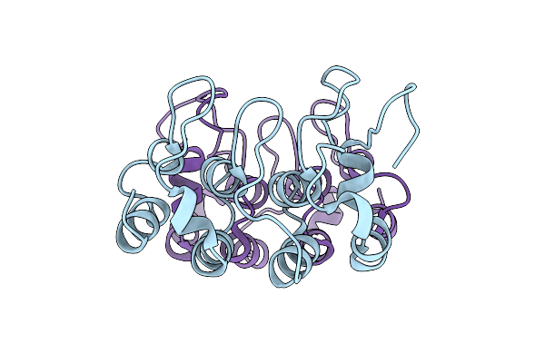
Crystal Structure Of Shikimate Kinase Of Mycobacterium Tuberculosis Complex With Shikimate-3-Phosphate
Organism: Mycobacterium tuberculosis h37rv
Method: X-RAY DIFFRACTION Resolution:1.73 Å Release Date: 2024-11-20 Classification: TRANSFERASE Ligands: S3P, SO4 |
 |
Organism: Mycobacterium tuberculosis
Method: X-RAY DIFFRACTION Resolution:1.75 Å Release Date: 2024-10-09 Classification: LYASE Ligands: MN, CL, PEG, EDO |
 |
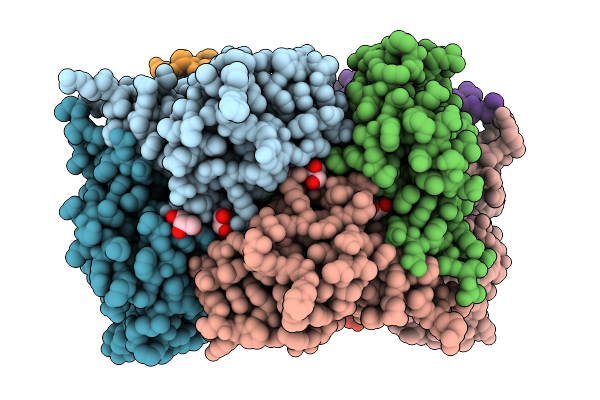
Crystal Structure Of The Ternary Complex Of Lactoperoxidase With Nitric Oxide And Nitrite Ion At 1.95 A Resolution
Organism: Bos taurus
Method: X-RAY DIFFRACTION Resolution:1.95 Å Release Date: 2024-09-11 Classification: OXIDOREDUCTASE Ligands: NAG, NO, NO2, NO3, SCN, IOD, HEM, EDO, CA, OSM |
 |
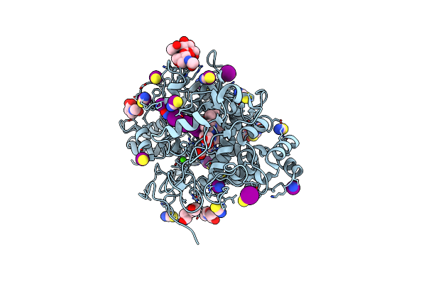
Structure Of Phosphopantetheine Adenylyltransferase (Ppat) From Enterobacter Spp. With The Expression Tag Bound In The Substrate Binding Site Of A Neighbouring Molecule At 2.39 A Resolution.
Organism: Enterobacter sp. 638
Method: X-RAY DIFFRACTION Resolution:2.39 Å Release Date: 2024-08-21 Classification: TRANSFERASE Ligands: PAE, GOL, EDO |
 |
Structure Of Phosphopantetheine Adenylyltransferase (Ppat) From Enterobacter Spp. With The Expression Tag Bound In The Substrate Binding Site Of A Neighbouring Molecule At 2.37 A Resolution.
Organism: Enterobacter sp. 638
Method: X-RAY DIFFRACTION Resolution:2.37 Å Release Date: 2024-08-21 Classification: TRANSFERASE Ligands: PAE, GOL, EDO |
 |
Structure Of Phosphopantetheine Adenylyltransferase (Ppat) From Enterobacter Spp. With The 17-Mer Expression Tag Bound In The Substrate Binding Site Of A Neighbouring Molecule At 2.60 A Resolution.
Organism: Enterobacter sp. 638
Method: X-RAY DIFFRACTION Resolution:2.60 Å Release Date: 2024-08-21 Classification: TRANSFERASE Ligands: CIT, GOL |