7AUA image
Deposition Date 2020-11-02
Release Date 2022-05-18
Last Version Date 2024-10-23
Entry Detail
PDB ID:
7AUA
Keywords:
Title:
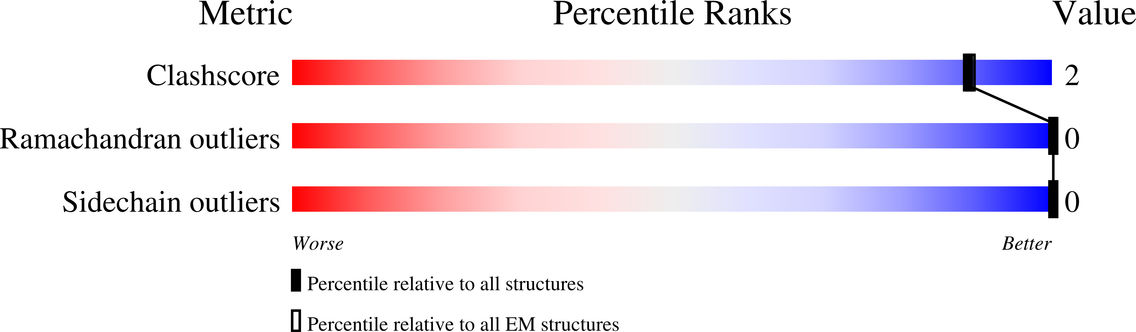
Cryo-EM structure of human exostosin-like 3 (EXTL3) in complex with UDP
Biological Source:
Source Organism:
Homo sapiens (Taxon ID: 9606)
Host Organism:
Method Details:
Experimental Method:
Resolution:
2.93 Å
Aggregation State:
PARTICLE
Reconstruction Method:
SINGLE PARTICLE
Macromolecular Entities
Polymer Type:polypeptide(L)
Molecule:Exostosin-like 3
Gene (Uniprot):EXTL3
Chain IDs:A, B
Chain Length:890
Number of Molecules:2
Biological Source:Homo sapiens
Primary Citation
The structure of EXTL3 helps to explain the different roles of bi-domain exostosins in heparan sulfate synthesis.
Nat Commun 13 3314 3314 (2022)
PMID: 35676258 DOI: 10.1038/s41467-022-31048-2

Abstact

Heparan sulfate is a highly modified O-linked glycan that performs diverse physiological roles in animal tissues. Though quickly modified, it is initially synthesised as a polysaccharide of alternating β-D-glucuronosyl and N-acetyl-α-D-glucosaminyl residues by exostosins. These enzymes generally possess two glycosyltransferase domains (GT47 and GT64)-each thought to add one type of monosaccharide unit to the backbone. Although previous structures of murine exostosin-like 2 (EXTL2) provide insight into the GT64 domain, the rest of the bi-domain architecture is yet to be characterised; hence, how the two domains co-operate is unknown. Here, we report the structure of human exostosin-like 3 (EXTL3) in apo and UDP-bound forms. We explain the ineffectiveness of EXTL3's GT47 domain to transfer β-D-glucuronosyl units, and we observe that, in general, the bi-domain architecture would preclude a processive mechanism of backbone extension. We therefore propose that heparan sulfate backbone polymerisation occurs by a simple dissociative mechanism.

Legend

Protein

Chemical

Disease

Primary Citation of related structures