4XCO image
Deposition Date 2014-12-18
Release Date 2015-06-10
Last Version Date 2024-01-10
Entry Detail
PDB ID:
4XCO
Title:
Signal-sequence induced conformational changes in the signal recognition particle
Biological Source:
Source Organism:
Host Organism:
Method Details:
Experimental Method:
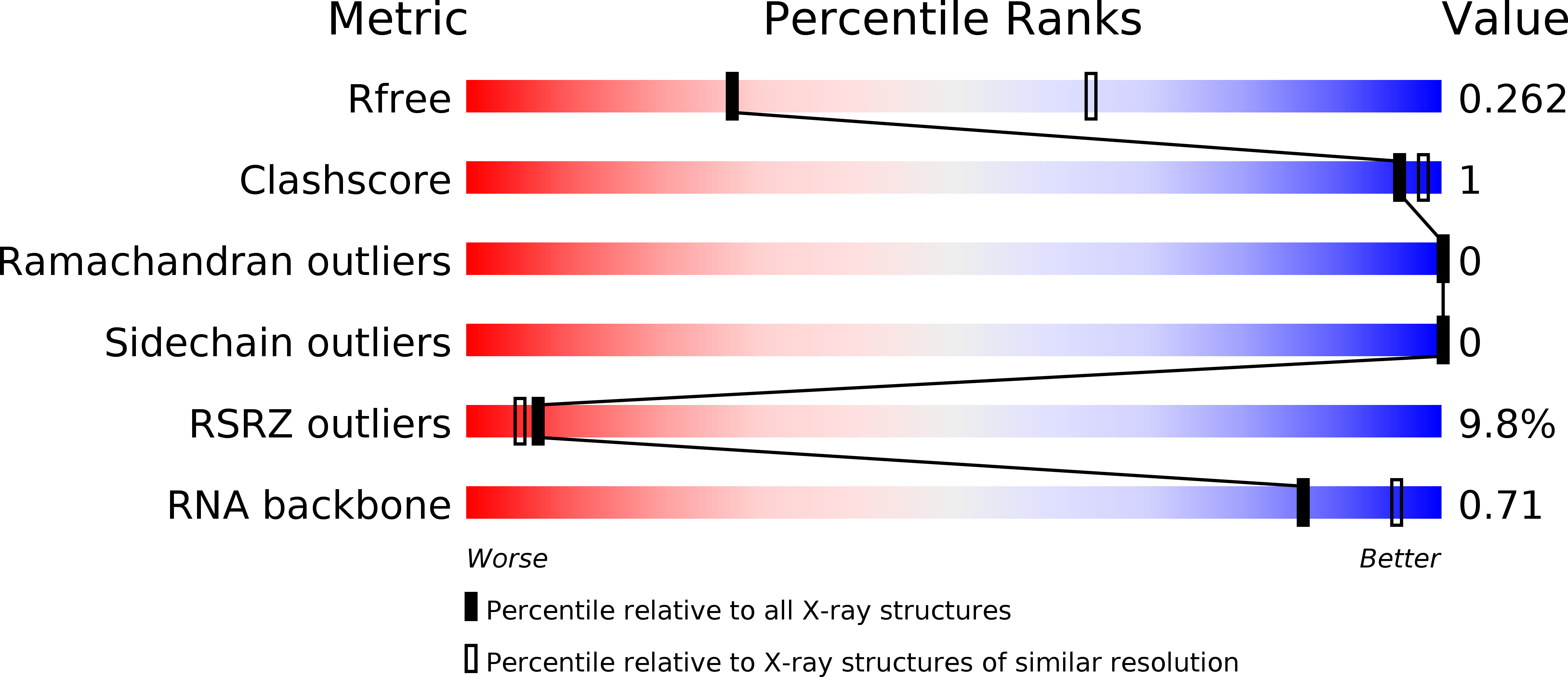
Resolution:
2.90 Å
R-Value Free:
0.25
R-Value Work:
0.20
R-Value Observed:
0.21
Space Group:
P 2 21 21
Macromolecular Entities
Structures with similar UniProt ID
Protein Blast
Polymer Type:polypeptide(L)
Molecule:Signal recognition particle 19 kDa protein
Gene (Uniprot):srp19
Chain IDs:B (auth: A), D (auth: B)
Chain Length:87
Number of Molecules:2
Biological Source:Methanocaldococcus jannaschii
Structures with similar UniProt ID
Protein Blast
Polymer Type:polypeptide(L)
Molecule:Signal recognition particle 54 kDa protein,signal sequence
Gene (Uniprot):srp54
Chain IDs:E (auth: C), F (auth: D)
Chain Length:173
Number of Molecules:2
Biological Source:Methanocaldococcus jannaschii
Polymer Type:polyribonucleotide
Molecule:RNA
Chain IDs:A (auth: M), C (auth: E)
Chain Length:96
Number of Molecules:2
Biological Source:Methanocaldococcus jannaschii
Primary Citation
Signal-sequence induced conformational changes in the signal recognition particle.
Nat Commun 6 7163 7163 (2015)
PMID: 26051119 DOI: 10.1038/ncomms8163

Abstact

Co-translational protein targeting is an essential, evolutionarily conserved pathway for delivering nascent proteins to the proper cellular membrane. In this pathway, the signal recognition particle (SRP) first recognizes the N-terminal signal sequence of nascent proteins and subsequently interacts with the SRP receptor. For this, signal sequence binding in the SRP54 M domain must be effectively communicated to the SRP54 NG domain that interacts with the receptor. Here we present the 2.9 Å crystal structure of unbound- and signal sequence bound SRP forms, both present in the asymmetric unit. The structures provide evidence for a coupled binding and folding mechanism in which signal sequence binding induces the concerted folding of the GM linker helix, the finger loop, and the C-terminal alpha helix αM6. This mechanism allows for a high degree of structural adaptability of the binding site and suggests how signal sequence binding in the M domain is coupled to repositioning of the NG domain.

Legend

Protein

Chemical

Disease

Primary Citation of related structures