4LVQ image
Deposition Date 2013-07-26
Release Date 2014-03-26
Last Version Date 2024-11-27
Entry Detail
PDB ID:
4LVQ
Title:
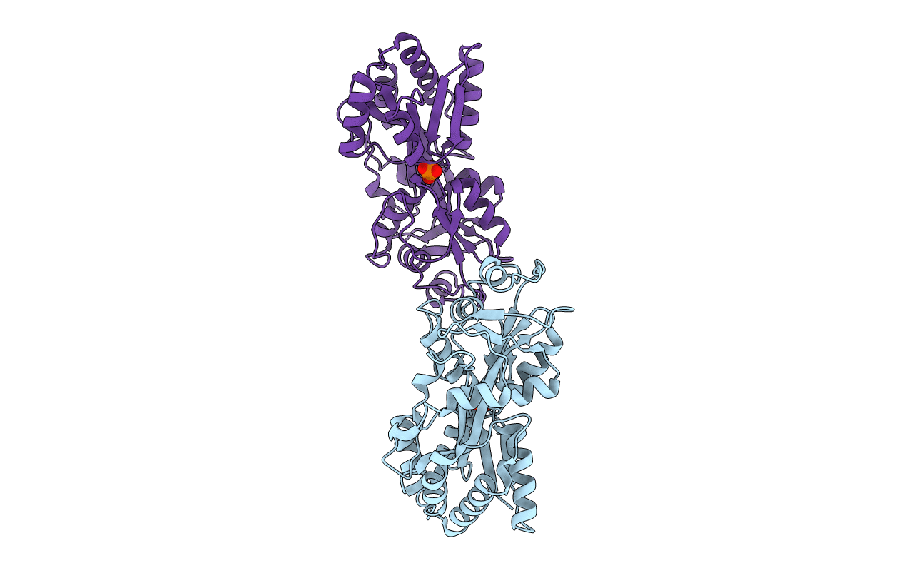
Crystal structure of the M. tuberculosis phosphate binding protein PstS3
Biological Source:
Source Organism:
Host Organism:
Method Details:
Experimental Method:
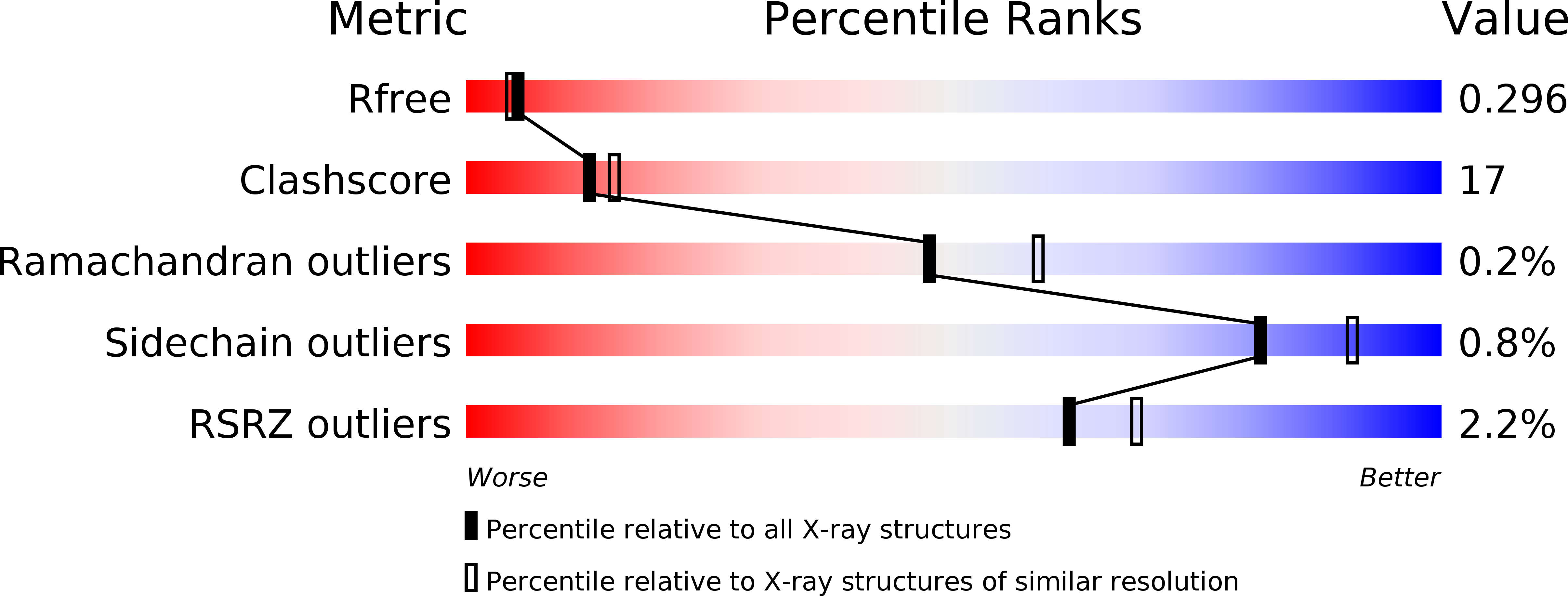
Resolution:
2.30 Å
R-Value Free:
0.27
R-Value Work:
0.23
R-Value Observed:
0.23
Space Group:
P 1 21 1
Macromolecular Entities
Structures with similar UniProt ID
Protein Blast
Polymer Type:polypeptide(L)
Molecule:Phosphate-binding protein PstS 3
Chain IDs:A, B
Chain Length:376
Number of Molecules:2
Biological Source:Mycobacterium tuberculosis
Ligand Molecules
Primary Citation
Crystal structure of the Mycobacterium tuberculosis phosphate binding protein PstS3.
Proteins 82 2268 2274 (2014)
PMID: 24615888 DOI: 10.1002/prot.24548

Abstact

Mycobacterium tuberculosis evades host immune responses by colonizing macrophages. Intraphagosomal M. tuberculosis is exposed to environmental stresses such as reactive oxygen and nitrogen intermediates as well as acid shock and inorganic phosphate (Pi) depletion. Experimental evidence suggests that expression levels of mycobacterial protein PstS3 (Rv0928) are significantly increased when M. tuberculosis bacilli are exposed to Pi starvation. Hence, PstS3 may be important for survival of Mtb in conditions where there is limited supply of Pi. We report here the structure of PstS3 from M. tuberculosis at 2.3-Å resolution. The protein presents a structure typical for ABC phosphate transfer receptors. Comparison with its cognate receptor PstS1 showed a different pattern distribution of surface charges in proximity to the Pi recognition site, suggesting complementary roles of the two proteins in Pi uptake.

Legend

Protein

Chemical

Disease

Primary Citation of related structures