Deposition Date
2008-08-20
Release Date
2009-07-28
Last Version Date
2024-11-06
Entry Detail
PDB ID:
3E8L
Keywords:
Title:
The Crystal Structure of the Double-headed Arrowhead Protease Inhibitor A in Complex with Two Trypsins
Biological Source:
Source Organism(s):
Sagittaria sagittifolia (Taxon ID: 4451)
Bos taurus (Taxon ID: 9913)
Bos taurus (Taxon ID: 9913)
Expression System(s):
Method Details:
Experimental Method:
Resolution:
2.48 Å
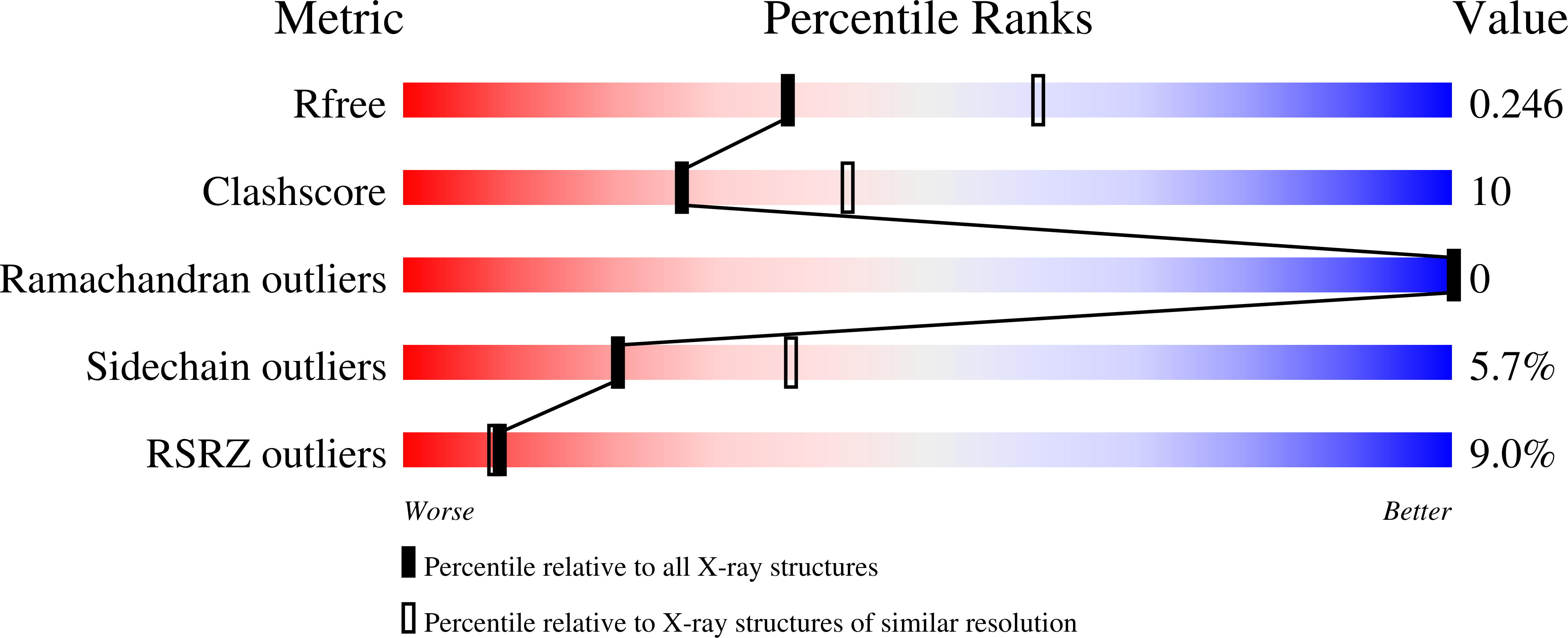
R-Value Free:
0.24
R-Value Work:
0.19
R-Value Observed:
0.19
Space Group:
C 2 2 21