1quq image
Deposition Date 1999-07-02
Release Date 1999-08-13
Last Version Date 2024-02-14
Entry Detail
PDB ID:
1QUQ
Title:
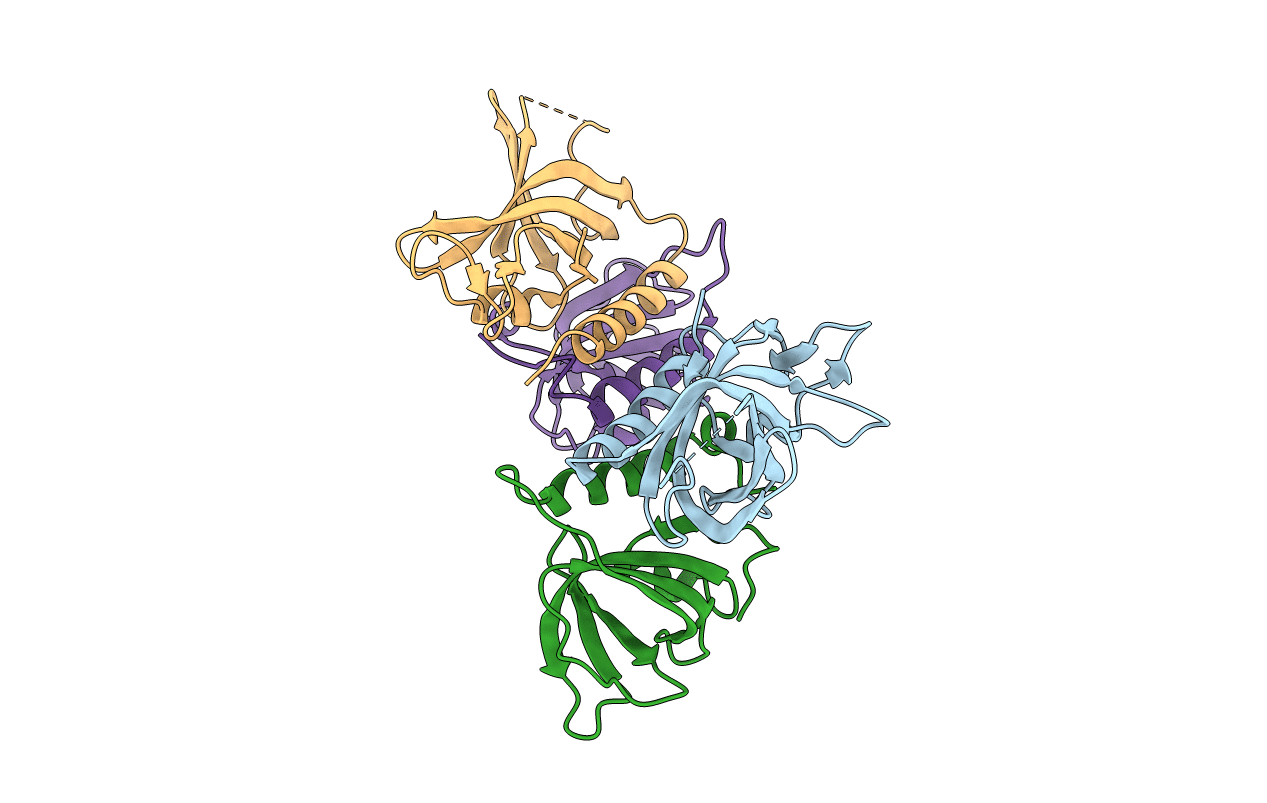
COMPLEX OF REPLICATION PROTEIN A SUBUNITS RPA14 AND RPA32
Biological Source:
Source Organism:
Homo sapiens (Taxon ID: 9606)
Host Organism:
Method Details:
Experimental Method:
Resolution:
2.50 Å
R-Value Free:
0.29
R-Value Work:
0.21
R-Value Observed:
0.21
Space Group:
P 21 21 21
Macromolecular Entities
Polymer Type:polypeptide(L)
Molecule:PROTEIN (REPLICATION PROTEIN A 32 KD SUBUNIT)
Gene (Uniprot):RPA2
Chain IDs:A, C
Chain Length:129
Number of Molecules:2
Biological Source:Homo sapiens
Polymer Type:polypeptide(L)
Molecule:PROTEIN (REPLICATION PROTEIN A 14 KD SUBUNIT)
Gene (Uniprot):RPA3
Chain IDs:B, D
Chain Length:121
Number of Molecules:2
Biological Source:Homo sapiens
Primary Citation
The crystal structure of the complex of replication protein A subunits RPA32 and RPA14 reveals a mechanism for single-stranded DNA binding.
EMBO J. 18 4498 4504 (1999)
PMID: 10449415 DOI: 10.1093/emboj/18.16.4498

Abstact

Replication protein A (RPA), the eukaryote single-stranded DNA-binding protein (SSB), is a heterotrimer. The largest subunit, RPA70, which harbours the major DNA-binding activity, has two DNA-binding domains that each adopt an OB-fold. The complex of the two smaller subunits, RPA32 and RPA14, has weak DNA-binding activity but the mechanism of DNA binding is unknown. We have determined the crystal structure of the proteolytic core of RPA32 and RPA14, which consists of the central two-thirds of RPA32 and the entire RPA14 subunit. The structure revealed that RPA14 and the central part of RPA32 are structural homologues. Each subunit contains a central OB-fold domain, which also resembles the DNA-binding domains in RPA70; an N-terminal extension that interacts with the central OB-fold domain; and a C-terminal helix that mediate heterodimerization via a helix-helix interaction. The OB-fold of RPA32, but not RPA14, possesses additional similarity to the RPA70 DNA-binding domains, supporting a DNA-binding role for RPA32. The discovery of a third and fourth OB-fold in RPA suggests that the quaternary structure of SSBs, which in Bacteria and Archaea are also tetramers of OB-folds, is conserved in evolution. The structure also suggests a mechanism for RPA trimer formation.

Legend

Protein

Chemical

Disease

Primary Citation of related structures