1RU0 image
Deposition Date 2003-12-10
Release Date 2004-10-12
Last Version Date 2023-08-23
Entry Detail
PDB ID:
1RU0
Keywords:
Title:
Crystal structure of DCoH2, a paralog of DCoH, the Dimerization Cofactor of HNF-1
Biological Source:
Source Organism:
Mus musculus (Taxon ID: 10090)
Method Details:
Experimental Method:
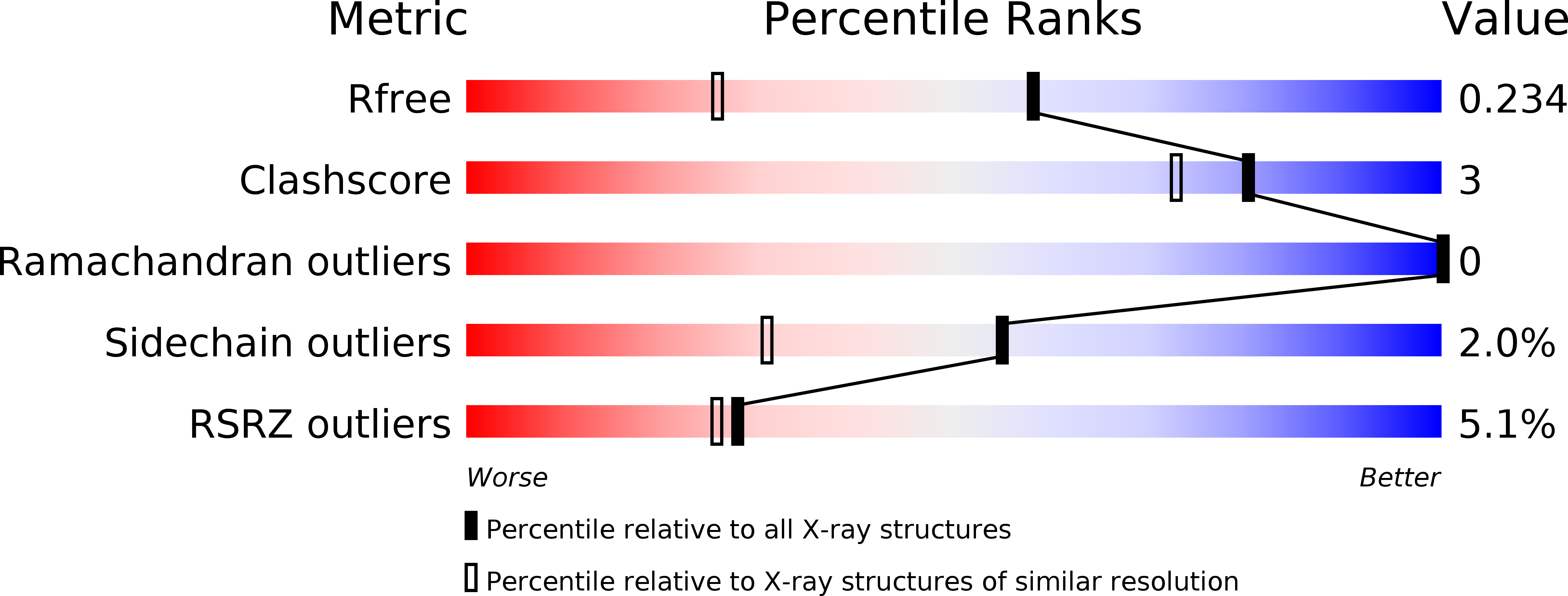
Resolution:
1.60 Å
R-Value Free:
0.23
R-Value Work:
0.21
R-Value Observed:
0.21
Space Group:
P 31 2 1
Macromolecular Entities
Structures with similar UniProt ID
Protein Blast
Polymer Type:polypeptide(L)
Molecule:DcoH-like protein DCoHm
Gene (Uniprot):Pcbd2
Chain IDs:A, B
Chain Length:105
Number of Molecules:2
Biological Source:Mus musculus
Primary Citation
Biochemical and structural basis for partially redundant enzymatic and transcriptional functions of DCoH and DCoH2
Biochemistry 43 7345 7355 (2004)
PMID: 15182178 DOI: 10.1021/bi049620t

Abstact

An inherited form of diabetes, maturity-onset diabetes of the young type 3 (MODY3), results from mutations in the transcriptional activator, hepatocyte nuclear factor-1alpha (HNF1alpha). Transcription by HNF1alpha is stimulated by the bifunctional coactivator DCoH (dimerization cofactor of HNF1). Strikingly, an HNF1alpha deletion in mice causes more severe phenotypes than a DCoH deletion. It has been hypothesized that a DCoH homolog, DCoH2, partially complements the DCoH deletion. To test this idea, we determined the biochemical properties and the 1.6-A-resolution crystal structure of DCoH2. Like DCoH, DCoH2 forms a tetramer, displays pterin-4alpha-carbinolamine dehydratase activity, and binds HNF1alpha in vivo and in vitro. DCoH and DCoH2 adopt identical folds with structural differences confined largely to the protein surfaces and the tetramer interface. In contrast to the hyperstable DCoH tetramer, DCoH2 readily disproportionates and forms a 2:2 complex with HNF1 in vitro. Phylogenetic analysis reveals six major subfamilies of DCoH proteins, including unique DCoH and DCoH2 branches in metazoans. These results suggest distinct roles for DCoH and DCoH2. Differences in conserved surface residues could mediate binding to different effectors. We propose that HNF1alpha binding kinetics may distinguish regulation by DCoH2, under thermodynamic control, from regulation by DCoH, under kinetic control.

Legend

Protein

Chemical

Disease

Primary Citation of related structures