1Q5X image
Deposition Date 2003-08-11
Release Date 2003-09-30
Last Version Date 2024-02-14
Entry Detail
PDB ID:
1Q5X
Title:
Structure of OF RRAA (MENG), a protein inhibitor of RNA processing
Biological Source:
Source Organism:
Escherichia coli (Taxon ID: 562)
Host Organism:
Method Details:
Experimental Method:
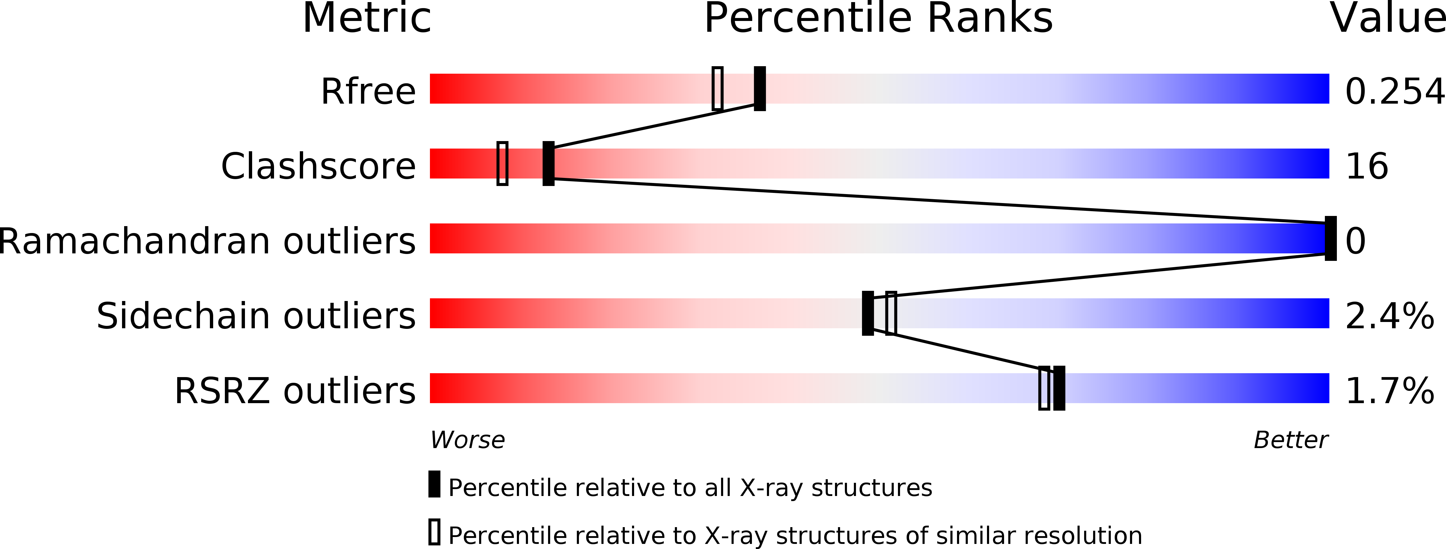
Resolution:
2.00 Å
R-Value Free:
0.27
R-Value Work:
0.22
R-Value Observed:
0.22
Space Group:
C 1 2 1
Macromolecular Entities
Structures with similar UniProt ID
Protein Blast
Polymer Type:polypeptide(L)
Molecule:REGULATOR OF RNASE E ACTIVITY A
Gene (Uniprot):rraA
Chain IDs:A, B, C
Chain Length:161
Number of Molecules:3
Biological Source:Escherichia coli
Primary Citation
The X-ray Structure of Escherichia coli RraA (MenG), A Protein Inhibitor of RNA Processing.
J.Mol.Biol. 332 1015 1024 (2003)
PMID: 14499605 DOI: 10.1016/S0022-2836(03)00970-7

Abstact

The Escherichia coli protein regulator of RNase E activity A (RraA) has recently been shown to act as a trans-acting modulator of RNA turnover in bacteria; it binds to the essential endonuclease RNase E and inhibits RNA processing in vivo and in vitro. Here, we report the 2.0A X-ray structure of RraA. The structure reveals a ring-like trimer with a central cavity of approximately 12A in diameter. Based on earlier sequence analysis, RraA had been identified as a putative S-adenosylmethionine:2-demethylmenaquinone and was annotated as MenG. However, an analysis of the RraA structure shows that the protein lacks the structural motifs usually required for methylases. Comparison of the observed fold with that of other proteins (and domains) suggests that the RraA fold is an ancient platform that has been adapted for a wide range of functions. An analysis of the amino acid sequence shows that the E.coli RraA exhibits an ancient relationship to a family of aldolases.

Legend

Protein

Chemical

Disease

Primary Citation of related structures