Deposition Date
2003-01-27
Release Date
2003-04-22
Last Version Date
2023-08-16
Entry Detail
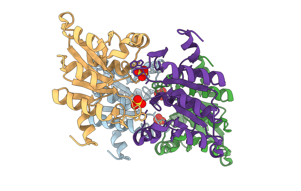
PDB ID:
1NRZ
Keywords:
Title:
Crystal structure of the IIBSor domain of the sorbose permease from Klebsiella pneumoniae solved to 1.75A resolution
Biological Source:
Source Organism:
Klebsiella pneumoniae (Taxon ID: 573)
Host Organism:
Method Details:
Experimental Method:
Resolution:
1.75 Å
R-Value Free:
0.24
R-Value Work:
0.21
R-Value Observed:
0.21
Space Group:
P 1 21 1