1DVK image
Deposition Date 2000-01-21
Release Date 2000-04-05
Last Version Date 2024-02-07
Entry Detail
PDB ID:
1DVK
Title:
CRYSTAL STRUCTURE OF THE FUNCTIONAL DOMAIN OF THE SPLICING FACTOR PRP18
Biological Source:
Source Organism:
Method Details:
Experimental Method:
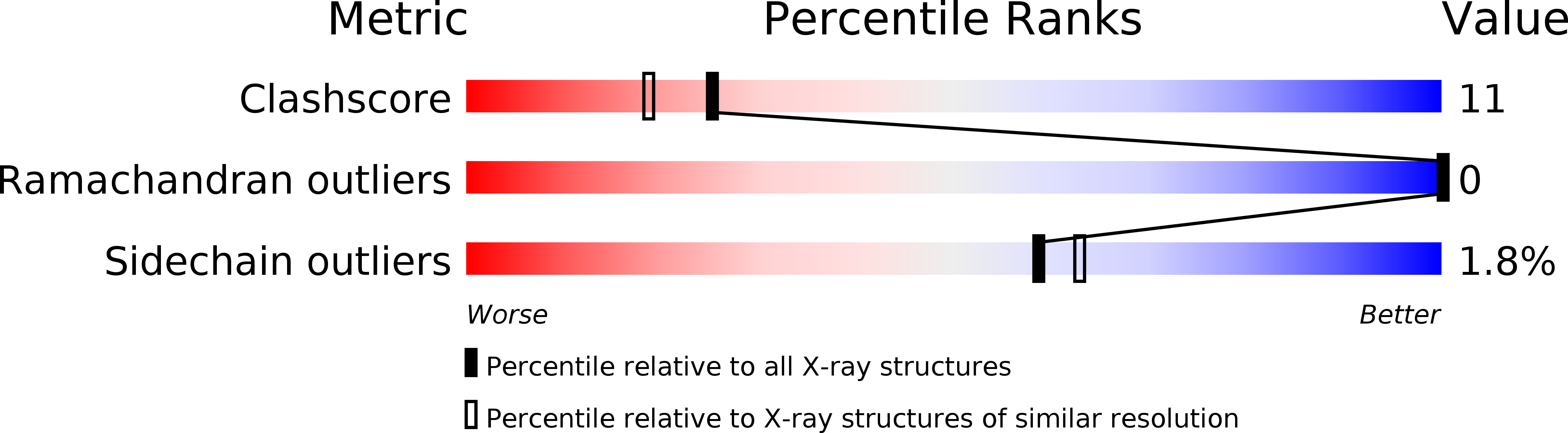
Resolution:
2.15 Å
R-Value Free:
0.25
R-Value Work:
0.20
R-Value Observed:
0.20
Space Group:
C 2 2 21
Macromolecular Entities
Structures with similar UniProt ID
Protein Blast
Polymer Type:polypeptide(L)
Molecule:PRP18
Gene (Uniprot):PRP18
Chain IDs:A, B
Chain Length:173
Number of Molecules:2
Biological Source:Saccharomyces cerevisiae
Primary Citation
Crystal structure of the functional domain of the splicing factor Prp18.
Proc.Natl.Acad.Sci.USA 97 3022 3027 (2000)
PMID: 10737784 DOI: 10.1073/pnas.97.7.3022

Abstact

The splicing factor Prp18 is required for the second step of pre-mRNA splicing. We have isolated and determined the crystal structure of a large fragment of the Saccharomyces cerevisiae Prp18 that lacks the N-terminal 79 amino acids. This fragment, called Prp18Delta79, is fully active in yeast splicing in vitro and includes the sequences of Prp18 that have been evolutionarily conserved. The core structure of Prp18Delta79 is compact and globular, consisting of five alpha-helices that adopt a novel fold that we have designated the five-helix X-bundle. The structure suggests that one face of Prp18 interacts with the splicing factor Slu7, whereas the more evolutionarily conserved amino acids in Prp18 form the opposite face. The most highly conserved region of Prp18, a nearly invariant stretch of 19 aa, forms part of a loop between two alpha-helices and may interact with the U5 small nuclear ribonucleoprotein particles. The structure is consistent with a model in which Prp18 forms a bridge between Slu7 and the U5 small nuclear ribonucleoprotein particles.

Legend

Protein

Chemical

Disease

Primary Citation of related structures