
Search Count: 10
 |
Organism: Sphaerobacter thermophilus (strain dsm 20745 / s 6022)
Method: X-RAY DIFFRACTION Resolution:2.10 Å Release Date: 2022-12-21 Classification: HYDROLASE |
 |
Crystal Structure Of Queuine Salvage Enzyme Duf2419 Complexed With Queuosine
Organism: Sphaerobacter thermophilus dsm 20745
Method: X-RAY DIFFRACTION Resolution:2.35 Å Release Date: 2022-12-21 Classification: HYDROLASE Ligands: QEO, MLI, PEG |
 |
Crystal Structure Of Queuine Salvage Enzyme Duf2419 Mutant K199C, Complexed With Queuosine
Organism: Sphaerobacter thermophilus dsm 20745
Method: X-RAY DIFFRACTION Resolution:2.50 Å Release Date: 2022-12-21 Classification: HYDROLASE Ligands: QEO, MLI |
 |
Crystal Structure Of Queuine Salvage Enzyme Duf2419, In Complex With Queuosine-5'-Monophosphate
Organism: Sphaerobacter thermophilus dsm 20745
Method: X-RAY DIFFRACTION Resolution:2.40 Å Release Date: 2022-12-21 Classification: HYDROLASE Ligands: 56B, NH4, PEG |
 |
Crystal Structure Of The Human Queuine Salvage Enzyme Duf2419, Wild-Type Apo Form
Organism: Homo sapiens
Method: X-RAY DIFFRACTION Resolution:1.78 Å Release Date: 2022-12-21 Classification: HYDROLASE Ligands: BTB |
 |
Crystal Structure Of Queuine Salvage Enzyme Duf2419, Wild-Type (Non-His6X Tagged)
Organism: Sphaerobacter thermophilus dsm 20745
Method: X-RAY DIFFRACTION Resolution:2.31 Å Release Date: 2022-12-21 Classification: HYDROLASE |
 |
Crystal Structure Of Queuine Salvage Enzyme Duf2419 Mutant D231N, In Complex With Queuosine-5'-Monophosphate
Organism: Sphaerobacter thermophilus dsm 20745
Method: X-RAY DIFFRACTION Resolution:1.99 Å Release Date: 2022-12-21 Classification: HYDROLASE Ligands: 56B, PEG |
 |
Crystal Structure Of The Human Queuine Salvage Enzyme Duf2419, Complexed With Queuine
Organism: Homo sapiens
Method: X-RAY DIFFRACTION Resolution:2.26 Å Release Date: 2022-12-21 Classification: HYDROLASE Ligands: QEI |
 |
Organism: Escherichia coli
Method: X-RAY DIFFRACTION Resolution:2.99 Å Release Date: 2009-04-28 Classification: MEMBRANE PROTEIN |
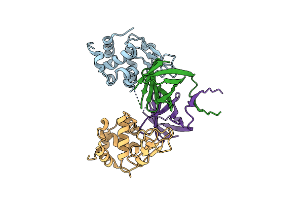 |
Crystal Structure Of Pseudomonas Aeruginosa Mlic In Complex With Hen Egg White Lysozyme
Organism: Pseudomonas aeruginosa, Gallus gallus
Method: X-RAY DIFFRACTION Resolution:2.50 Å Release Date: 2008-12-23 Classification: HYDROLASE |