Search Count: 17
 |
Organism: Chaetomium thermophilum var. thermophilum dsm 1495
Method: X-RAY DIFFRACTION Resolution:2.57 Å Release Date: 2017-06-28 Classification: PROTEIN TRANSPORT |
 |
Organism: Mus musculus, Saccharomyces cerevisiae
Method: ELECTRON MICROSCOPY Release Date: 2017-06-28 Classification: TRANSPORT PROTEIN |
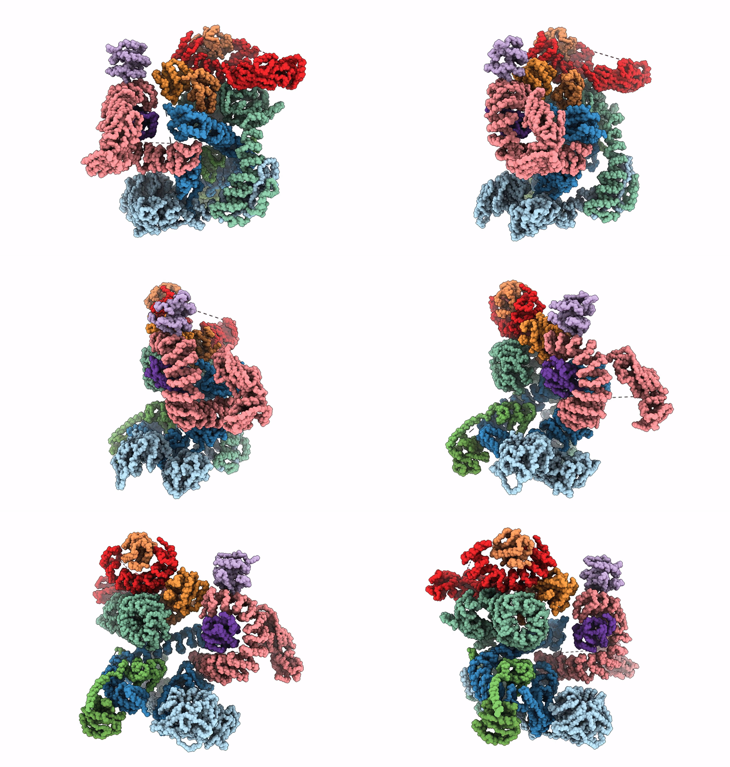 |
The Structure Of The Copi Coat Leaf In Complex With The Arfgap2 Uncoating Factor
Organism: Mus musculus, Saccharomyces cerevisiae, Rattus norvegicus
Method: ELECTRON MICROSCOPY Release Date: 2017-06-28 Classification: TRANSPORT PROTEIN |
 |
Organism: Mus musculus, Saccharomyces cerevisiae
Method: ELECTRON MICROSCOPY Release Date: 2017-06-28 Classification: TRANSPORT PROTEIN |
 |
Organism: Mus musculus, Saccharomyces cerevisiae
Method: ELECTRON MICROSCOPY Release Date: 2017-06-28 Classification: TRANSPORT PROTEIN |
 |
Organism: Mus musculus, Saccharomyces cerevisiae
Method: ELECTRON MICROSCOPY Release Date: 2017-06-28 Classification: TRANSPORT PROTEIN |
 |
Organism: Saccharomyces cerevisiae, Mus musculus
Method: ELECTRON MICROSCOPY Resolution:13.00 Å Release Date: 2015-07-08 Classification: TRANSPORT PROTEIN |
 |
Organism: Saccharomyces cerevisiae, Mus musculus
Method: ELECTRON MICROSCOPY Resolution:21.00 Å Release Date: 2015-07-08 Classification: TRANSPORT PROTEIN |
 |
Organism: Saccharomyces cerevisiae, Mus musculus
Method: ELECTRON MICROSCOPY Resolution:18.00 Å Release Date: 2015-07-08 Classification: TRANSPORT PROTEIN |
 |
Organism: Saccharomyces cerevisiae, Mus musculus
Method: ELECTRON MICROSCOPY Resolution:23.00 Å Release Date: 2015-07-08 Classification: TRANSPORT PROTEIN |
 |
Organism: Saccharomyces cerevisiae, Mus musculus
Method: ELECTRON MICROSCOPY Resolution:21.00 Å Release Date: 2015-07-08 Classification: TRANSPORT PROTEIN |
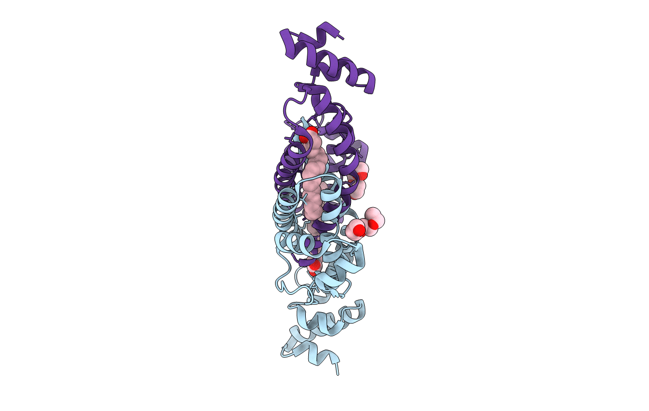 |
Structure Of Rv1264N, The Regulatory Domain Of The Mycobacterial Adenylyl Cylcase Rv1264, At Ph 6.0
Organism: Mycobacterium tuberculosis
Method: X-RAY DIFFRACTION Resolution:1.60 Å Release Date: 2006-11-07 Classification: LYASE Ligands: OLA, 1PE |
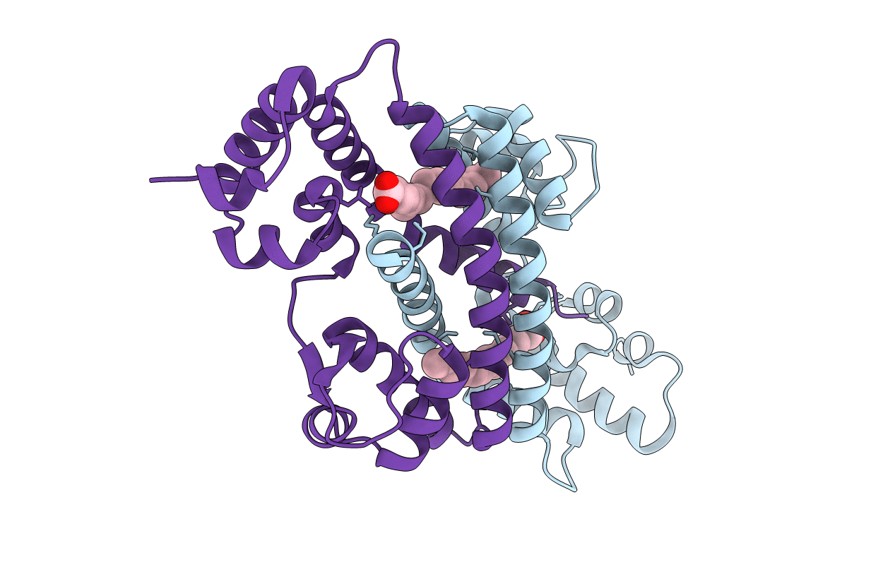 |
Structure Of Rv1264N, The Regulatory Domain Of The Mycobacterial Adenylyl Cylcase Rv1264, At Ph 8.5
Organism: Mycobacterium tuberculosis
Method: X-RAY DIFFRACTION Resolution:2.35 Å Release Date: 2006-11-07 Classification: LYASE Ligands: OLA |
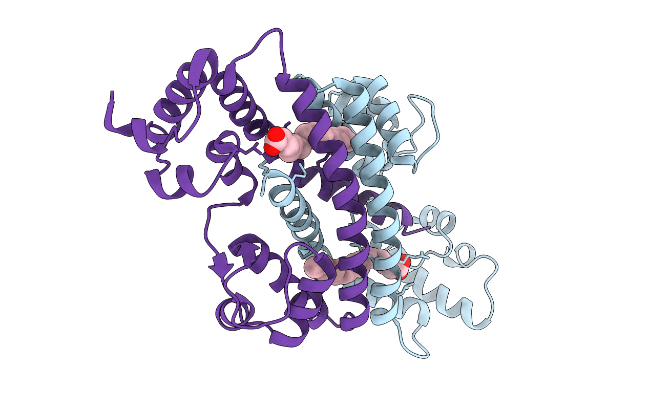 |
Structure Of Rv1264N, The Regulatory Domain Of The Mycobacterial Adenylyl Cylcase Rv1264, At Ph 5.3
Organism: Mycobacterium tuberculosis
Method: X-RAY DIFFRACTION Resolution:2.68 Å Release Date: 2006-11-07 Classification: LYASE Ligands: OLA |
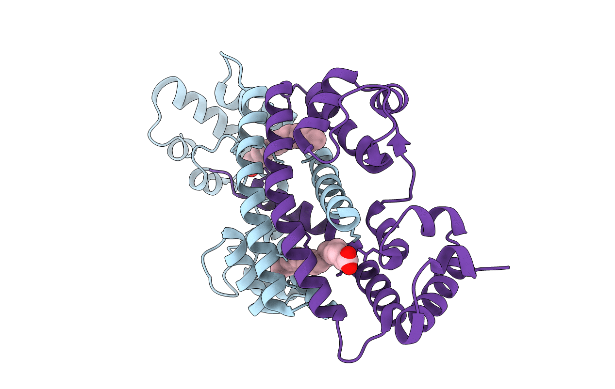 |
Structure Of Rv1264N, The Regulatory Domain Of The Mycobacterial Adenylyl Cylcase Rv1264, With A Salt Precipitant
Organism: Mycobacterium tuberculosis
Method: X-RAY DIFFRACTION Resolution:2.28 Å Release Date: 2006-11-07 Classification: LYASE Ligands: CL, OLA |
 |
Structure Of The Dimerized Cytoplasmic Domain Of P23 In Solution, Nmr, 10 Structures
|
 |

