
Search Count: 15
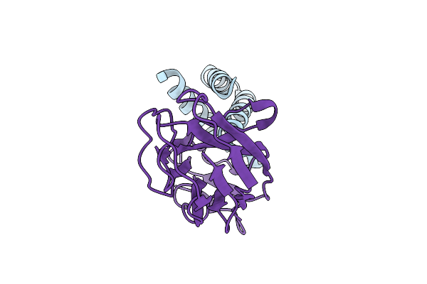 |
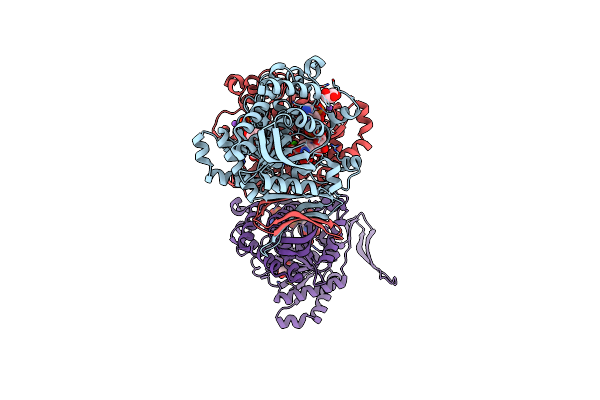
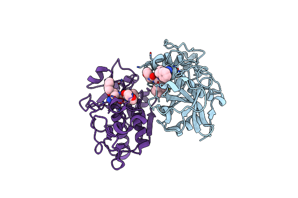
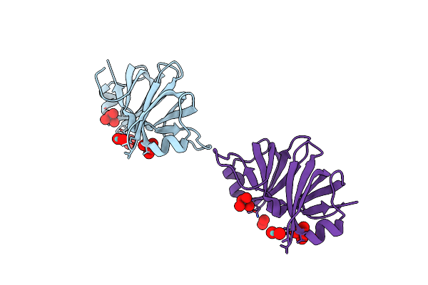
Organism: Synthetic construct, Acinetobacter baumannii acicu
Method: X-RAY DIFFRACTION Release Date: 2025-09-17 Classification: PROTEIN BINDING |
 |
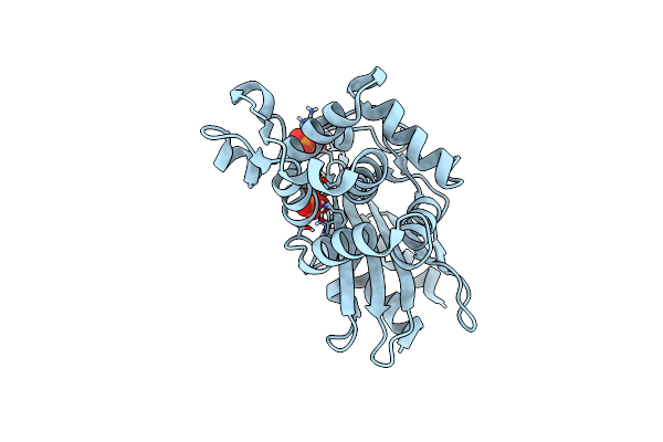
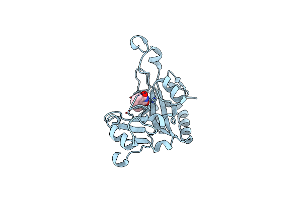
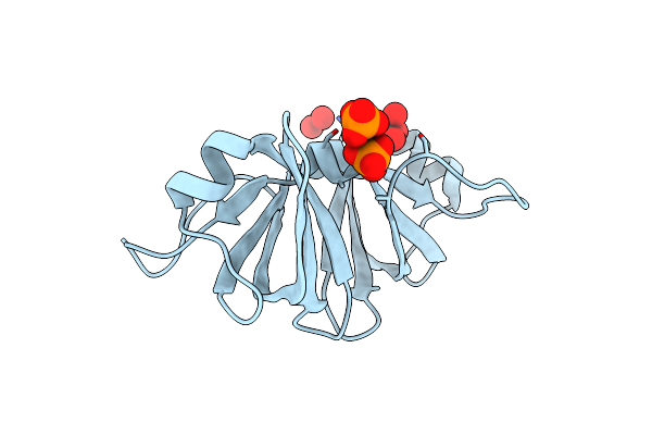
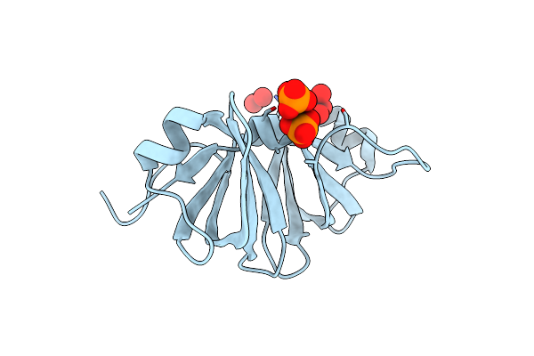
Organism: Synthetic construct, Escherichia coli
Method: X-RAY DIFFRACTION Release Date: 2025-09-17 Classification: DE NOVO PROTEIN |
 |
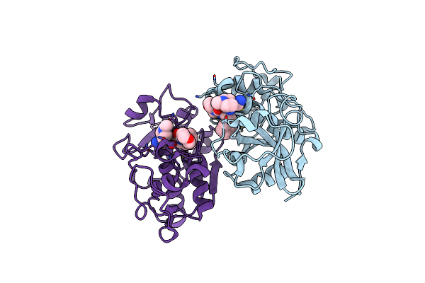
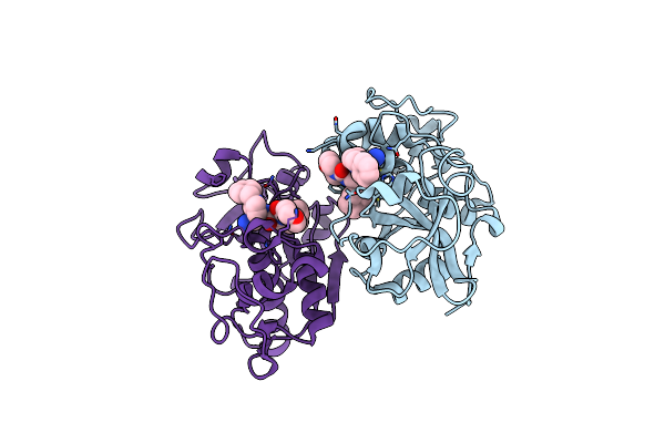
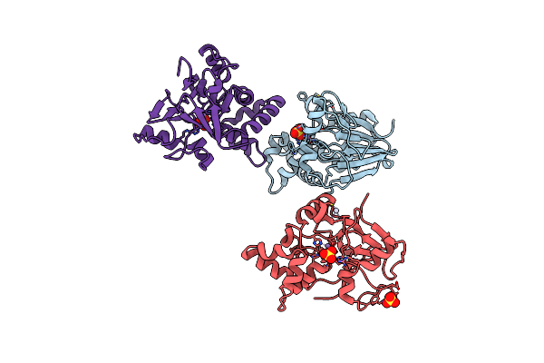
Phosphoserine Blac, Class A Serine Beta-Lactamase From Mycobacterium Tuberculosis
Organism: Mycobacterium tuberculosis
Method: X-RAY DIFFRACTION Resolution:1.52 Å Release Date: 2019-08-28 Classification: HYDROLASE Ligands: PO4 |
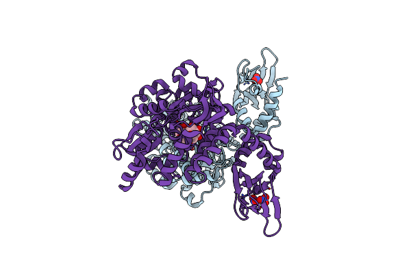 |
Organism: Escherichia coli o157:h7, Mus musculus
Method: X-RAY DIFFRACTION Resolution:2.04 Å Release Date: 2016-11-30 Classification: TRANSPORT PROTEIN Ligands: NO3 |
 |
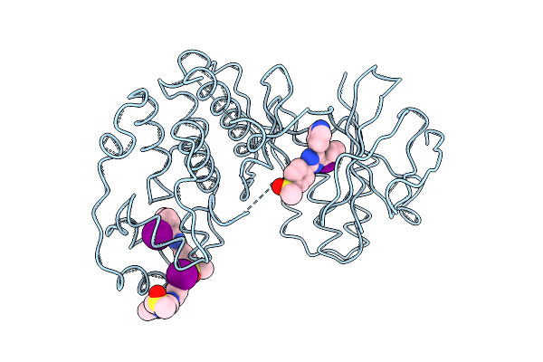
A Novel Phospho-Switch In The Linker Region Of The Snail Zinc Finger Protein Which Regulates 14-3-3 Association, Dna Binding And Epithelial-Mesenchymal Differentiation
Organism: Homo sapiens
Method: X-RAY DIFFRACTION Resolution:1.45 Å Release Date: 2015-06-17 Classification: SIGNALING PROTEIN Ligands: MG, GOL |
 |
Crystal Structure Of Human Cytosolic Nadp(+)-Dependent Isocitrate Dehydrogenase R132H Mutant In Complex With Nadph, Alpha-Ketoglutarate And Calcium(2+)
Organism: Homo sapiens
Method: X-RAY DIFFRACTION Resolution:2.10 Å Release Date: 2009-11-24 Classification: OXIDOREDUCTASE Ligands: NDP, AKG, CA, NA, GOL |
 |
Catalytic Fragment Of Cholix Toxin From Vibrio Cholerae In Complex With The 1,8-Naphthalimide Inhibitor
Organism: Vibrio cholerae
Method: X-RAY DIFFRACTION Resolution:1.19 Å Release Date: 2009-09-15 Classification: TRANSFERASE,TOXIN Ligands: 18N |
 |
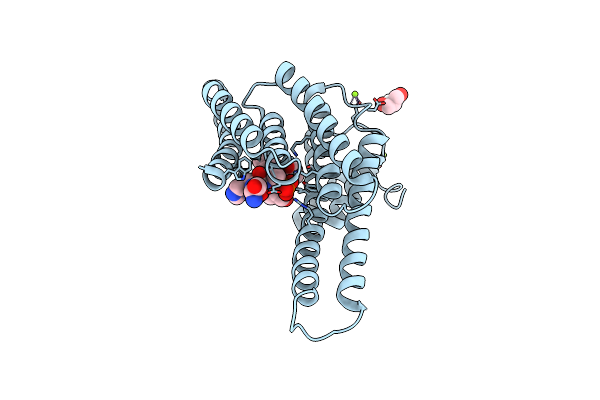
Organism: Homo sapiens
Method: X-RAY DIFFRACTION Resolution:1.97 Å Release Date: 2007-12-18 Classification: HYDROLASE Ligands: Y11 |
 |
Organism: Homo sapiens
Method: X-RAY DIFFRACTION Resolution:2.00 Å Release Date: 2007-12-18 Classification: HYDROLASE Ligands: Y14 |
 |
Organism: Homo sapiens
Method: X-RAY DIFFRACTION Resolution:2.00 Å Release Date: 2007-12-18 Classification: HYDROLASE Ligands: Y15 |
 |
Two Crystal Structures Of The Cytoplasmic Molybdate-Binding Protein Modg Suggest A Novel Cooperative Binding Mechanism And Provide Insights Into Ligand-Binding Specificity. Phosphate-Grown Form With Molybdate And Phosphate Bound
Organism: Azotobacter vinelandii
Method: X-RAY DIFFRACTION Resolution:1.80 Å Release Date: 2001-05-11 Classification: BINDING PROTEIN Ligands: MOO, PO4 |
 |
Two Crystal Structures Of The Cytoplasmic Molybdate-Binding Protein Modg Suggest A Novel Cooperative Binding Mechanism And Provide Insights Into Ligand-Binding Specificity. Phosphate-Grown Form With Tungstate And Phosphate Bound
Organism: Azotobacter vinelandii
Method: X-RAY DIFFRACTION Resolution:1.80 Å Release Date: 2001-05-11 Classification: BINDING PROTEIN Ligands: WO4, PO4 |
 |
Two Crystal Structures Of The Cytoplasmic Molybdate-Binding Protein Modg Suggest A Novel Cooperative Binding Mechanism And Provide Insights Into Ligand-Binding Specificity. Peg-Grown Form With Molybdate Bound
Organism: Azotobacter vinelandii
Method: X-RAY DIFFRACTION Resolution:1.65 Å Release Date: 2001-05-11 Classification: BINDING PROTEIN Ligands: MOO |
 |
Organism: Enterobacteria phage lambda
Method: X-RAY DIFFRACTION Resolution:2.15 Å Release Date: 2001-03-07 Classification: Viral protein, hydrolase Ligands: MN, HG, SO4 |
 |
Organism: Homo sapiens
Method: X-RAY DIFFRACTION Resolution:2.00 Å Release Date: 1998-05-06 Classification: SERINE/THREONINE-PROTEIN KINASE Ligands: D13 |