
Search Count: 10
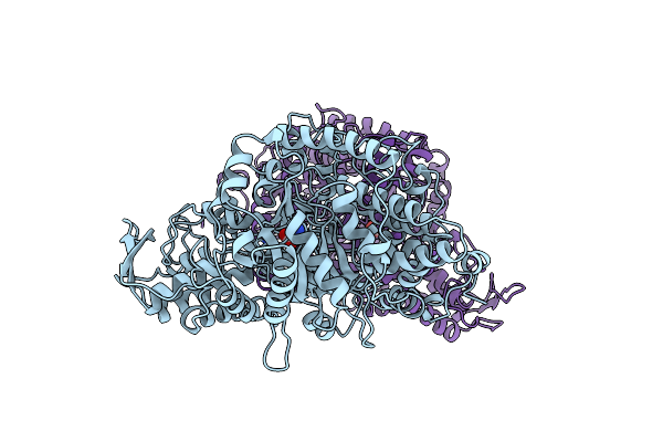 |
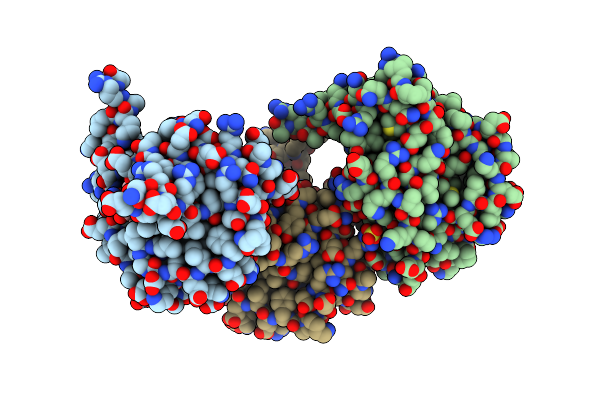
Crystal Structure Of Pfl From E.Coli In Complex With Substrate Analogue Oxamate
Organism: Escherichia coli
Method: X-RAY DIFFRACTION Resolution:2.60 Å Release Date: 2000-05-31 Classification: LYASE/TRANSFERASE Ligands: OXM |
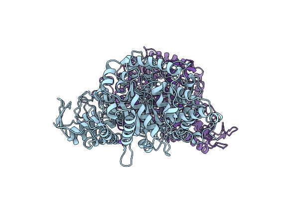 |
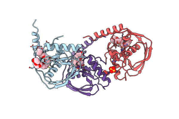
Organism: Escherichia coli
Method: X-RAY DIFFRACTION Resolution:2.90 Å Release Date: 1999-12-15 Classification: LYASE Ligands: CL, NA |
 |
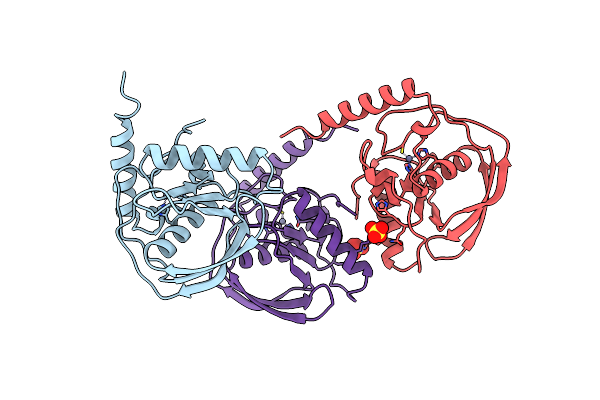
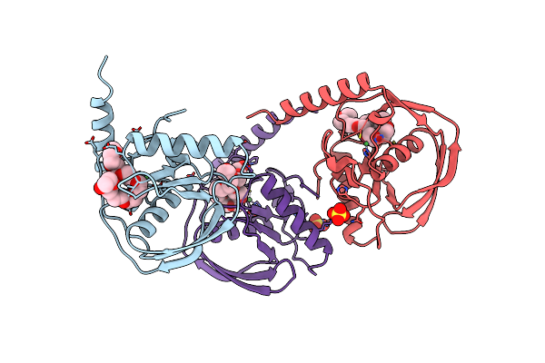
Organism: Escherichia coli
Method: X-RAY DIFFRACTION Resolution:2.30 Å Release Date: 1999-12-08 Classification: TRANSFERASE Ligands: CO3, NA |
 |
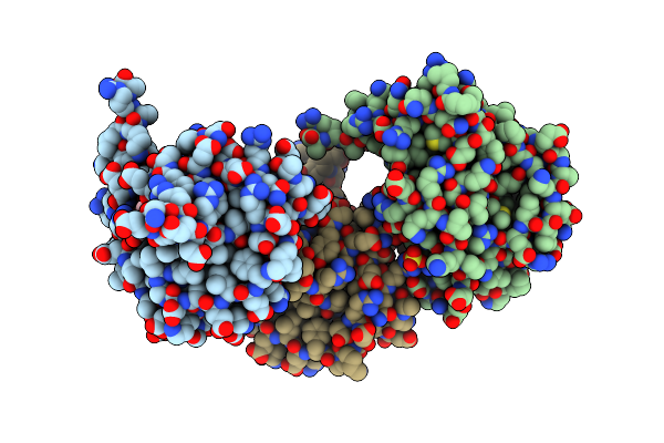
Peptide Deformylase As Zn2+ Containing Form (Native) In Complex With Inhibitor Polyethylene Glycol
Organism: Escherichia coli
Method: X-RAY DIFFRACTION Resolution:1.90 Å Release Date: 1999-08-27 Classification: HYDROLASE Ligands: ZN, 2PE, SO4 |
 |
Organism: Escherichia coli
Method: X-RAY DIFFRACTION Resolution:2.50 Å Release Date: 1999-08-27 Classification: HYDROLASE Ligands: ZN, SO4 |
 |
Peptide Deformylase As Ni2+ Containing Form In Complex With Tripeptide Met-Ala-Ser
Organism: Escherichia coli
Method: X-RAY DIFFRACTION Resolution:2.10 Å Release Date: 1999-08-27 Classification: HYDROLASE Ligands: NI, SO4 |
 |
Organism: Escherichia coli
Method: X-RAY DIFFRACTION Resolution:2.50 Å Release Date: 1999-08-27 Classification: HYDROLASE Ligands: NI, SO4 |
 |
Peptide Deformylase As Zn2+ Containing Form In Complex With Tripeptide Met-Ala-Ser
Organism: Escherichia coli
Method: X-RAY DIFFRACTION Resolution:2.20 Å Release Date: 1999-08-27 Classification: HYDROLASE Ligands: ZN, SO4 |
 |
Peptide Deformylase As Fe2+ Containing Form (Native) In Complex With Inhibitor Polyethylene Glycol
Organism: Escherichia coli
Method: X-RAY DIFFRACTION Resolution:1.90 Å Release Date: 1999-08-26 Classification: HYDROLASE Ligands: FE, 2PE, SO4 |
 |
Pdf Protein Is Crystallized As Ni2+ Containing Form, Cocrystallized With Inhibitor Polyethylene Glycol (Peg)
Organism: Escherichia coli
Method: X-RAY DIFFRACTION Resolution:1.90 Å Release Date: 1999-03-23 Classification: HYDROLASE Ligands: NI, 2PE, SO4 |