
Search Count: 2,227
 |
Organism: Stichodactyla helianthus
Method: ELECTRON MICROSCOPY Release Date: 2025-10-29 Classification: MEMBRANE PROTEIN Ligands: FO4, CLR |
 |
Organism: Actinia fragacea
Method: ELECTRON MICROSCOPY Release Date: 2025-10-29 Classification: MEMBRANE PROTEIN Ligands: FO4, CLR |
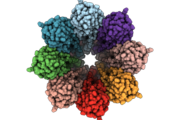 |
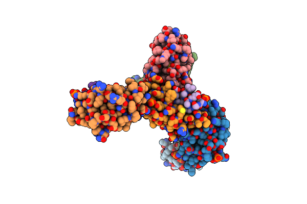
Structure Of The Octameric Pore Of Fragaceotxin C (Frac Or Delta-Actitoxin-Afr1A) In Large Unilamellar Vesicles.
Organism: Actinia fragacea
Method: ELECTRON MICROSCOPY Release Date: 2025-10-08 Classification: MEMBRANE PROTEIN Ligands: FO4, CLR |
 |
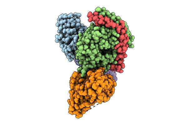
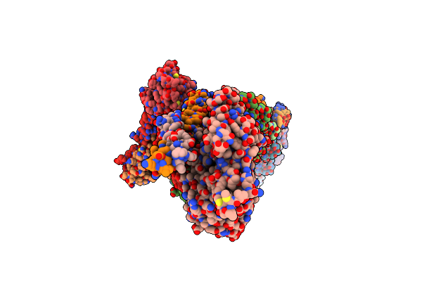
Structure Of 6Mer Pore Intermediate Of Sticholysin Ii (Stnii) Toxin In Lipid Nanodiscs
Organism: Stichodactyla helianthus
Method: ELECTRON MICROSCOPY Release Date: 2025-10-08 Classification: MEMBRANE PROTEIN Ligands: FO4 |
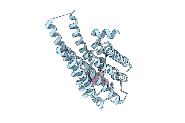 |
Local Refinement Of Drd2 Bound To Lsd In Complex With A Mini-Goa And Scfv16 Obtained By Cryo-Electron Microscopy (Cryoem)
Organism: Escherichia coli, Homo sapiens
Method: ELECTRON MICROSCOPY Release Date: 2025-09-17 Classification: MEMBRANE PROTEIN Ligands: 7LD |
 |
Global Reconstruction Of Drd2 Bound To Lsd In Complex With A Mini-Goa And Scfv16 Obtained By Cryo-Electron Microscopy (Cryoem)
Organism: Homo sapiens, Escherichia coli, Mus musculus
Method: ELECTRON MICROSCOPY Release Date: 2025-09-17 Classification: MEMBRANE PROTEIN Ligands: 7LD |
 |
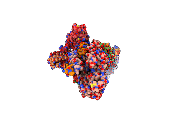
Structure Of 5Mer Pore Intermediate Of Sticholysin Ii (Stnii) Toxin In Lipid Nanodiscs
Organism: Stichodactyla helianthus
Method: ELECTRON MICROSCOPY Release Date: 2025-09-17 Classification: MEMBRANE PROTEIN Ligands: FO4 |
 |
Organism: Rhinolophus cornutus, Sarbecovirus
Method: ELECTRON MICROSCOPY Release Date: 2025-08-27 Classification: VIRAL PROTEIN |
 |
Organism: Homo sapiens
Method: ELECTRON MICROSCOPY Release Date: 2025-08-27 Classification: VIRAL PROTEIN Ligands: NAG |
 |
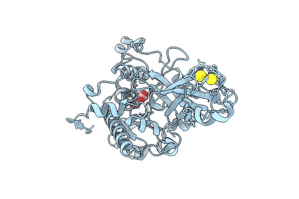
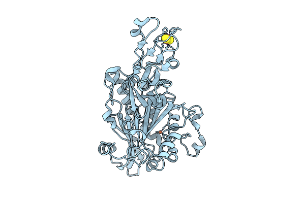
Co-Crystal Structure Of Human 8-Oxoguanine Glycosylase N149C Mutant With Dna Containing Photocaged 8-Oxoguanine
Organism: Homo sapiens
Method: X-RAY DIFFRACTION Release Date: 2025-07-23 Classification: LYASE Ligands: CA, GOL |
 |
Co-Crystal Structure Of Human 8-Oxoguanine Glycosylase N149C Mutant With Dna Containing Photocaged 8-Oxoguanine After Deprotection
Organism: Homo sapiens
Method: X-RAY DIFFRACTION Release Date: 2025-07-23 Classification: LYASE Ligands: CA, GOL |
 |
Organism: Comamonas testosteroni kf-1
Method: X-RAY DIFFRACTION Release Date: 2025-07-16 Classification: OXIDOREDUCTASE Ligands: FES, FE2, 8G0 |
 |
Crystal Structure Of Isophthalate Dioxygenase From Comamonas Testosteroni Kf1
Organism: Comamonas testosteroni kf-1
Method: X-RAY DIFFRACTION Release Date: 2025-07-09 Classification: OXIDOREDUCTASE Ligands: FE2, FES |
 |
Crystal Structure Of Hdm2 In Complex With A Peptidic Ligand Containing A Di-Urea Insert.
Organism: Homo sapiens, Synthetic construct
Method: X-RAY DIFFRACTION Release Date: 2025-07-02 Classification: ANTITUMOR PROTEIN Ligands: PO4 |
 |
Organism: Homo sapiens, Bos taurus
Method: X-RAY DIFFRACTION Release Date: 2025-06-11 Classification: TRANSFERASE/INHIBITOR Ligands: A1L62 |
 |
Structure Of Native Murine Cardiac Thin Filament At Pca=5.8 In Ca2+-Free Rotated State (Upper Strand)
Organism: Mus musculus
Method: ELECTRON MICROSCOPY Release Date: 2025-06-11 Classification: MOTOR PROTEIN Ligands: ADP, MG |
 |
Structure Of Native Murine Cardiac Thin Filament At Pca=5.8 In Ca2+-Free Tilted State (Upper Strand)
Organism: Mus musculus
Method: ELECTRON MICROSCOPY Release Date: 2025-06-11 Classification: MOTOR PROTEIN Ligands: ADP, MG |
 |
Structure Of Native Murine Cardiac Thin Filament At Pca=5.8 In Ca2+-Bound Partially Activated State (Upper Strand)
Organism: Mus musculus
Method: ELECTRON MICROSCOPY Release Date: 2025-06-11 Classification: MOTOR PROTEIN Ligands: ADP, MG, CA |
 |
Structure Of Native Murine Cardiac Thin Filament At Pca=5.8 In Ca2+-Bound Fully Activated State (Upper Strand)
Organism: Mus musculus
Method: ELECTRON MICROSCOPY Release Date: 2025-06-11 Classification: MOTOR PROTEIN Ligands: ADP, MG, CA |
 |
Structure Of Native Murine Cardiac Thin Filament Variant I79N In Troponin T At Pca=5.8 In Ca2+-Free State (Lower Strand)
Organism: Mus musculus
Method: ELECTRON MICROSCOPY Release Date: 2025-06-11 Classification: MOTOR PROTEIN Ligands: ADP, MG |