
Search Count: 12
 |
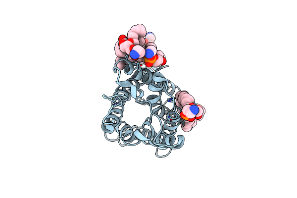
Crystal Structure Of The Receptor Binding Domain Of Sars-Cov-2 Spike Protein In Complex With Ce9
Organism: Severe acute respiratory syndrome coronavirus 2, Synthetic construct
Method: X-RAY DIFFRACTION Release Date: 2025-01-15 Classification: VIRAL PROTEIN/INHIBITOR Ligands: NAG, GOL |
 |
Crystal Structure Of The Receptor Binding Domain Of Sars-Cov-2 Omicron Ba.2 Variant Spike Protein In Complex With Ce149
Organism: Severe acute respiratory syndrome coronavirus 2, Synthetic construct
Method: X-RAY DIFFRACTION Release Date: 2025-01-15 Classification: VIRAL PROTEIN/INHIBITOR Ligands: GOL |
 |
Crystal Structure Of The Receptor Binding Domain Of Sars-Cov-2 Alpha Variant Spike Protein In Complex With Ce59
Organism: Severe acute respiratory syndrome coronavirus 2, Synthetic construct
Method: X-RAY DIFFRACTION Release Date: 2025-01-15 Classification: VIRAL PROTEIN/INHIBITOR Ligands: NAG |
 |
Crystal Structure Of The Receptor Binding Domain Of Sars-Cov-2 Delta Variant Spike Protein In Complex With Ce59
Organism: Severe acute respiratory syndrome coronavirus 2, Synthetic construct
Method: X-RAY DIFFRACTION Release Date: 2025-01-15 Classification: VIRAL PROTEIN/INHIBITOR Ligands: GOL |
 |
Crystal Structure Of The Receptor Binding Domain Of Sars-Cov-2 Alpha Variant Spike Protein In Complex With Ce41
Organism: Severe acute respiratory syndrome coronavirus 2, Synthetic construct
Method: X-RAY DIFFRACTION Release Date: 2025-01-15 Classification: VIRAL PROTEIN/INHIBITOR Ligands: NAG |
 |
Crystal Structure Of The Receptor Binding Domain Of Sars-Cov-2 Omicron Ba.2 Variant Spike Protein In Complex With Cespiace
Organism: Severe acute respiratory syndrome coronavirus 2, Synthetic construct
Method: X-RAY DIFFRACTION Release Date: 2025-01-15 Classification: VIRAL PROTEIN/INHIBITOR Ligands: GOL |
 |
Crystal Structure Of The Receptor Binding Domain Of Sars-Cov-2 Omicron Ba.5 Variant Spike Protein In Complex With Cespiace
Organism: Severe acute respiratory syndrome coronavirus 2, Synthetic construct
Method: X-RAY DIFFRACTION Release Date: 2025-01-15 Classification: VIRAL PROTEIN/INHIBITOR Ligands: GOL |
 |
Crystal Structure Of The Receptor Binding Domain Of Sars-Cov-2 Omicron Xbb.1.5 Variant Spike Protein In Complex With Cespiace
Organism: Severe acute respiratory syndrome coronavirus 2, Synthetic construct
Method: X-RAY DIFFRACTION Release Date: 2025-01-15 Classification: VIRAL PROTEIN/INHIBITOR Ligands: GOL, NA |
 |
Cryo-Em Structure Of Sars-Cov-2 Spike Ectodomain (Hexapro, Omicron Ba.2 Variant) In Complex With Cespiace
Organism: Severe acute respiratory syndrome coronavirus 2, Synthetic construct
Method: ELECTRON MICROSCOPY Release Date: 2025-01-15 Classification: VIRAL PROTEIN/INHIBITOR Ligands: NAG |
 |
Cryo-Em Structure Of Sars-Cov-2 Spike Ectodomain (Hexapro, Omicron Ba.5 Variant) In Complex With Cespiace, Class 1
Organism: Severe acute respiratory syndrome coronavirus 2, Synthetic construct
Method: ELECTRON MICROSCOPY Release Date: 2025-01-15 Classification: VIRAL PROTEIN/INHIBITOR Ligands: NAG |
 |
Cryo-Em Structure Of Sars-Cov-2 Spike Ectodomain (Hexapro, Omicron Ba.5 Variant) In Complex With Cespiace, Class 2
Organism: Severe acute respiratory syndrome coronavirus 2, Synthetic construct
Method: ELECTRON MICROSCOPY Release Date: 2025-01-15 Classification: VIRAL PROTEIN/INHIBITOR Ligands: NAG |
 |
Structure Of Aquaporin-4 S180D Mutant At 2.8 A Resolution By Electron Crystallography
Organism: Rattus norvegicus
Method: ELECTRON CRYSTALLOGRAPHY Resolution:2.80 Å Release Date: 2009-06-09 Classification: TRANSPORT PROTEIN Ligands: PEE |