
Search Count: 20
 |
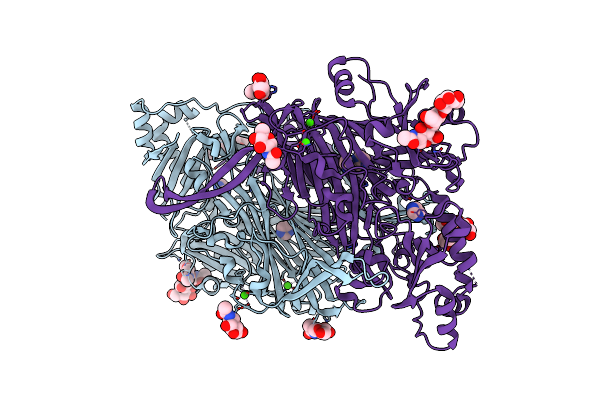
The Crystal Structure Of M644G Variant Of Dna Pol Epsilon Containing Dttp In The Polymerase Active Site
Organism: Saccharomyces cerevisiae, Synthetic construct
Method: X-RAY DIFFRACTION Resolution:2.60 Å Release Date: 2023-11-01 Classification: DNA BINDING PROTEIN Ligands: CA, TTP, ACT |
 |
The Crystal Structure Of M644G Variant Of Dna Pol Epsilon Containing Ctp In The Polymerase Active Site
Organism: Saccharomyces cerevisiae, Synthetic construct
Method: X-RAY DIFFRACTION Resolution:2.60 Å Release Date: 2023-10-25 Classification: DNA BINDING PROTEIN Ligands: CA, CTP, ACT |
 |
The Crystal Structure Of M644G Variant Of Dna Pol Epsilon Containing Dctp In The Polymerase Active Site
Organism: Saccharomyces cerevisiae, Synthetic construct
Method: X-RAY DIFFRACTION Resolution:2.50 Å Release Date: 2023-10-25 Classification: DNA BINDING PROTEIN Ligands: CA, DCP |
 |
The Crystal Structure Of N828V Variant Of Dna Pol Epsilon Containing Datp In The Polymerase Active Site
Organism: Saccharomyces cerevisiae, Synthetic construct
Method: X-RAY DIFFRACTION Resolution:2.70 Å Release Date: 2023-10-25 Classification: DNA BINDING PROTEIN Ligands: DTP, CA |
 |
The Crystal Structure Of M644G Variant Of Dna Pol Epsilon Containing Utp In The Polymerase Active Site
Organism: Saccharomyces cerevisiae, Synthetic construct
Method: X-RAY DIFFRACTION Resolution:2.65 Å Release Date: 2023-10-25 Classification: DNA BINDING PROTEIN Ligands: UTP, CA, ACT |
 |
The Crystal Structure Of N828V Variant Of Dna Pol Epsilon Containing Utp In The Polymerase Active Site
Organism: Saccharomyces cerevisiae, Synthetic construct
Method: X-RAY DIFFRACTION Resolution:2.60 Å Release Date: 2023-10-25 Classification: DNA BINDING PROTEIN Ligands: UTP, CA, GOL |
 |
The Crystal Structure Of The L439V Variant Of Pol2Core In Complex With Dna And An Incoming Nucleotide
Organism: Saccharomyces cerevisiae, Synthetic construct
Method: X-RAY DIFFRACTION Resolution:2.46 Å Release Date: 2022-07-27 Classification: DNA BINDING PROTEIN Ligands: DTP, CA, ACT |
 |
The Crystal Structure Of The V426L Variant Of Pol2Core In Complex With Dna And An Incoming Nucleotide
Organism: Saccharomyces cerevisiae, Synthetic construct
Method: X-RAY DIFFRACTION Resolution:2.60 Å Release Date: 2022-07-27 Classification: DNA BINDING PROTEIN Ligands: DTP, CA |
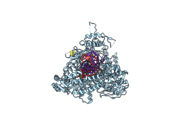 |
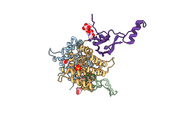
The Crystal Structure Of Pol2Core In Complex With Dna And An Incoming Nucleotide, Carrying An Fe-S Cluster
Organism: Saccharomyces cerevisiae (strain atcc 204508 / s288c), Synthetic construct
Method: X-RAY DIFFRACTION Resolution:2.70 Å Release Date: 2019-04-24 Classification: DNA BINDING PROTEIN Ligands: SF4, DTP, MG |
 |
The Crystal Structure Of Pol2Core In Complex With Dna And An Incoming Nucleotide, Carrying An Fe-S Cluster
Organism: Saccharomyces cerevisiae, Synthetic construct
Method: X-RAY DIFFRACTION Resolution:2.80 Å Release Date: 2019-04-17 Classification: DNA BINDING PROTEIN Ligands: SF4, DTP, CA |
 |
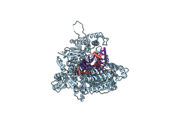
The Crystal Structure Of Pol2Core-M644G In Complex With Dna And An Incoming Nucleotide
Organism: Saccharomyces cerevisiae (strain atcc 204508 / s288c), Synthetic construct
Method: X-RAY DIFFRACTION Resolution:2.50 Å Release Date: 2019-01-30 Classification: DNA BINDING PROTEIN Ligands: DTP, CA, FE |
 |
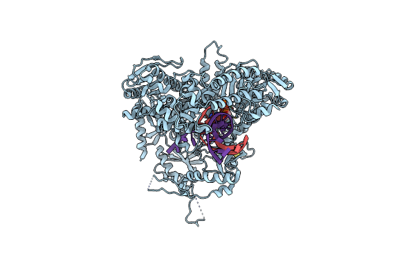
The Crystal Structure Of The Pol2 Catalytic Domain Of Dna Polymerase Epsilon Carrying A P301R Substitution.
Organism: Saccharomyces cerevisiae (strain atcc 204508 / s288c), Synthetic construct
Method: X-RAY DIFFRACTION Resolution:2.62 Å Release Date: 2019-01-30 Classification: DNA BINDING PROTEIN Ligands: DTP, CA, FE |
 |
The Crystal Structure Of The Pol2 Catalytic Domain Of Dna Polymerase Epsilon Carrying A P301R Substitution.
Organism: Saccharomyces cerevisiae (strain atcc 204508 / s288c), Synthetic construct
Method: X-RAY DIFFRACTION Resolution:2.65 Å Release Date: 2019-01-30 Classification: DNA BINDING PROTEIN Ligands: DTP, CA, FE |
 |
Organism: Homo sapiens
Method: X-RAY DIFFRACTION Resolution:1.90 Å Release Date: 2011-10-26 Classification: CELL ADHESION Ligands: MG |
 |
Organism: Homo sapiens
Method: X-RAY DIFFRACTION Resolution:2.60 Å Release Date: 2011-06-15 Classification: OXIDOREDUCTASE Ligands: CU, CA, IMD, NAG, FMT |
 |
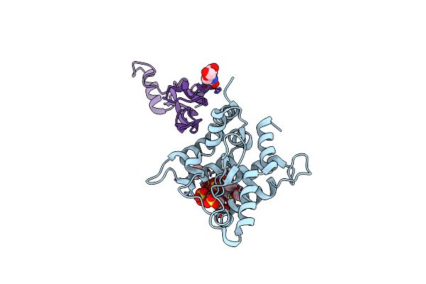
The Crystal Structure Of Human Soluble Primary Amine Oxidase Aoc3 In The Off-Copper Conformation
Organism: Homo sapiens
Method: X-RAY DIFFRACTION Resolution:2.95 Å Release Date: 2011-06-15 Classification: OXIDOREDUCTASE Ligands: CU, CA, IMD, NAG, MAN |
 |
Organism: Homo sapiens, Rattus norvegicus
Method: X-RAY DIFFRACTION Resolution:2.35 Å Release Date: 2009-06-02 Classification: HORMONE Ligands: EDO, NAG, SO4 |
 |
Organism: Homo sapiens
Method: X-RAY DIFFRACTION Resolution:1.60 Å Release Date: 2009-03-17 Classification: HORMONE Ligands: PO4 |
 |
Organism: Homo sapiens
Method: X-RAY DIFFRACTION Resolution:2.80 Å Release Date: 2009-03-17 Classification: HORMONE |
 |
The Structure Of The Gdnf:Coreceptor Complex: Insights Into Ret Signalling And Heparin Binding.
Organism: Rattus norvegicus, Homo sapiens
Method: X-RAY DIFFRACTION Resolution:2.35 Å Release Date: 2008-10-21 Classification: RECEPTOR/GLYCOPROTEIN COMPLEX Ligands: EDO, NAG |