
Search Count: 13
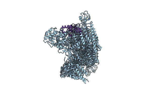 |
Organism: Mus musculus, Homo sapiens
Method: ELECTRON MICROSCOPY Release Date: 2022-06-01 Classification: SIGNALING PROTEIN Ligands: ATP, MG, ZN |
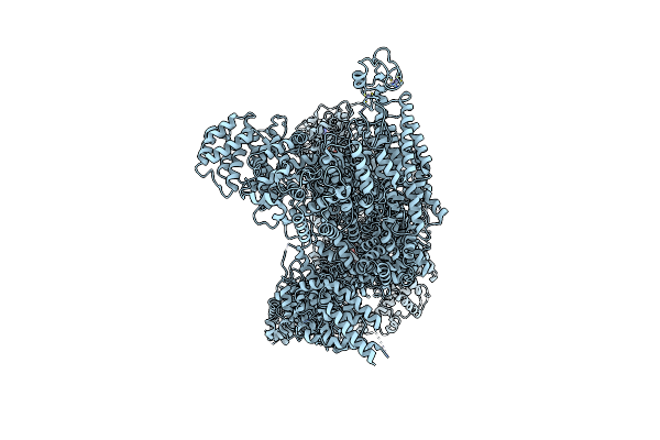 |
Organism: Mus musculus
Method: ELECTRON MICROSCOPY Release Date: 2022-06-01 Classification: SIGNALING PROTEIN Ligands: ATP, MG, ZN, ADP, AGS |
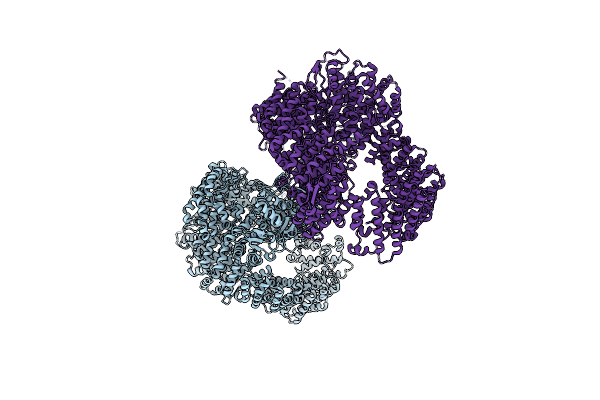 |
Organism: Nematocida sp. ertm5
Method: X-RAY DIFFRACTION Resolution:3.04 Å Release Date: 2021-07-28 Classification: LIGASE |
 |
Organism: Mus musculus
Method: ELECTRON MICROSCOPY Release Date: 2020-07-01 Classification: SIGNALING PROTEIN Ligands: ATP, MG, ZN |
 |
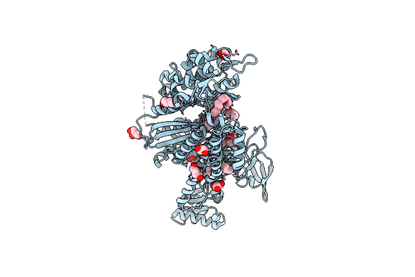
Mouse Rnf213 Mutant R4753K Modeling The Moyamoya-Disease-Related Human Variant R4810K
Organism: Mus musculus
Method: ELECTRON MICROSCOPY Release Date: 2020-07-01 Classification: SIGNALING PROTEIN Ligands: ATP, MG, ZN |
 |
Molecular Features Of The Unc-45 Chaperone Critical For Binding And Folding Muscle Myosin
Organism: Caenorhabditis elegans
Method: X-RAY DIFFRACTION Resolution:1.88 Å Release Date: 2019-10-30 Classification: MOTOR PROTEIN Ligands: ADP, GOL, BU1, PGO, HEZ, P15 |
 |
Molecular Features Of The Unc-45 Chaperone Critical For Binding And Folding Muscle Myosin
Organism: Caenorhabditis elegans
Method: X-RAY DIFFRACTION Resolution:3.40 Å Release Date: 2019-10-30 Classification: CHAPERONE |
 |
Molecular Features Of The Unc-45 Chaperone Critical For Binding And Folding Muscle Myosin
Organism: Caenorhabditis elegans
Method: X-RAY DIFFRACTION Resolution:2.93 Å Release Date: 2019-10-30 Classification: CHAPERONE |
 |
Molecular Features Of The Unc-45 Chaperone Critical For Binding And Folding Muscle Myosin
Organism: Caenorhabditis elegans
Method: X-RAY DIFFRACTION Resolution:3.80 Å Release Date: 2019-10-30 Classification: CHAPERONE |
 |
Crystal Structure Of The Myosin Chaperone Unc-45 From C. Elegans (Alternative Conformation)
Organism: Caenorhabditis elegans
Method: X-RAY DIFFRACTION Resolution:3.80 Å Release Date: 2018-02-14 Classification: CHAPERONE |
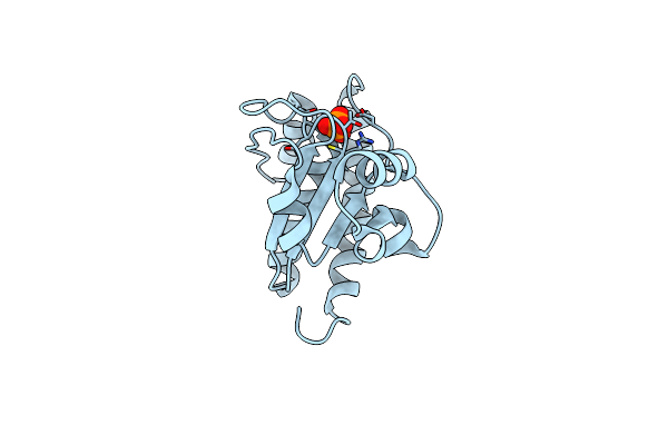 |
Organism: Bacillus subtilis subsp. subtilis
Method: X-RAY DIFFRACTION Resolution:1.70 Å Release Date: 2013-07-03 Classification: HYDROLASE Ligands: PO4 |
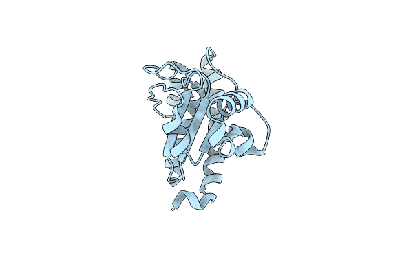 |
Ywle Arginine Phosphatase - C7S Mutant With Phosphorylated Active Site Serine
Organism: Bacillus subtilis subsp. subtilis
Method: X-RAY DIFFRACTION Resolution:1.80 Å Release Date: 2013-07-03 Classification: HYDROLASE |
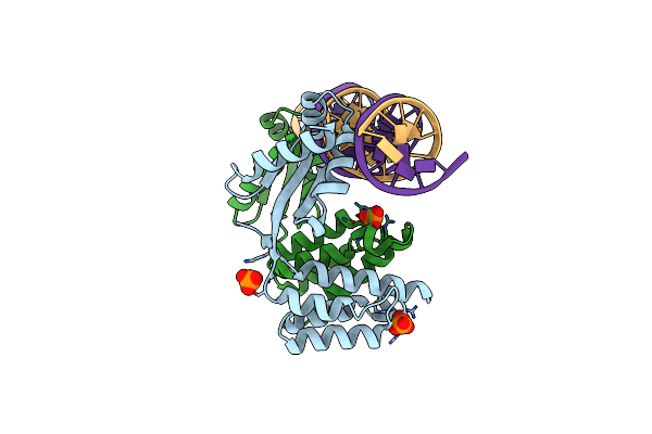 |
Organism: Bacillus stearothermophilus
Method: X-RAY DIFFRACTION Resolution:2.40 Å Release Date: 2009-06-30 Classification: TRANSCRIPTION/DNA Ligands: PO4 |