
Search Count: 27
 |
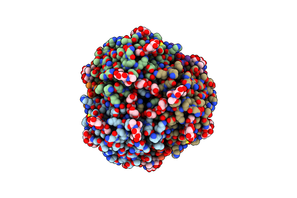
Organism: Lassa virus josiah
Method: ELECTRON MICROSCOPY Release Date: 2025-09-17 Classification: VIRAL PROTEIN Ligands: NAG, UNX |
 |
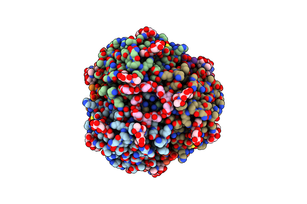
Organism: Lassa virus josiah, Homo sapiens
Method: ELECTRON MICROSCOPY Release Date: 2025-08-27 Classification: VIRAL PROTEIN Ligands: NAG, UNX, A1BK9 |
 |
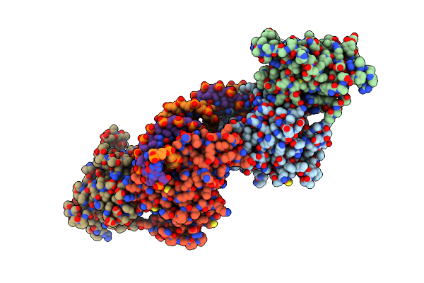
Organism: Lassa virus josiah, Homo sapiens
Method: ELECTRON MICROSCOPY Release Date: 2025-08-27 Classification: VIRAL PROTEIN Ligands: NAG, UNX |
 |
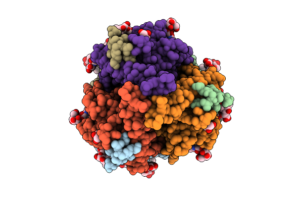
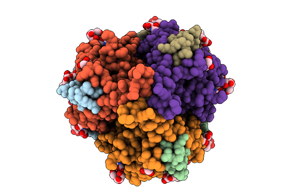
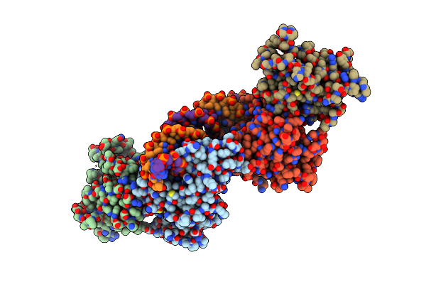
Focused Reconstruction Of The Transmembrane Region Of The Lassa Virus Spike Complex
Organism: Lassa virus josiah
Method: ELECTRON MICROSCOPY Release Date: 2025-08-27 Classification: VIRAL PROTEIN |
 |
Organism: Lassa virus josiah, Homo sapiens
Method: ELECTRON MICROSCOPY Release Date: 2025-08-27 Classification: VIRAL PROTEIN Ligands: NAG, UNX |
 |
Organism: Lassa virus josiah
Method: ELECTRON MICROSCOPY Release Date: 2025-08-27 Classification: VIRAL PROTEIN Ligands: NAG, UNX |
 |
Organism: Homo sapiens
Method: ELECTRON MICROSCOPY Release Date: 2025-07-02 Classification: VIRAL PROTEIN Ligands: NAG |
 |
Organism: Sabia virus
Method: ELECTRON MICROSCOPY Release Date: 2025-06-11 Classification: VIRAL PROTEIN Ligands: NAG, ZN |
 |
Organism: Sabia virus
Method: ELECTRON MICROSCOPY Release Date: 2025-06-11 Classification: VIRAL PROTEIN Ligands: NAG, K |
 |
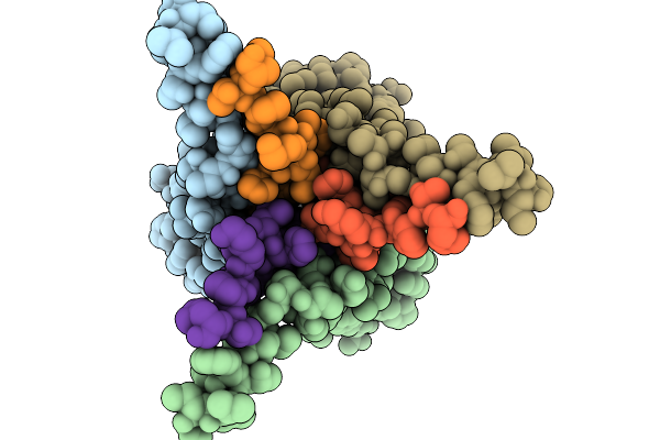
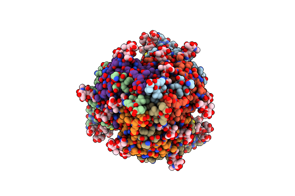
Structure Of The Sabia Virus Spike Complex H157M Mutant In A Closed Conformation
Organism: Sabia virus
Method: ELECTRON MICROSCOPY Release Date: 2025-06-11 Classification: VIRAL PROTEIN Ligands: NAG, ZN |
 |
Organism: Listeria seeligeri serovar 1/2b str. slcc3954
Method: ELECTRON MICROSCOPY Release Date: 2025-04-23 Classification: RNA BINDING PROTEIN/RNA |
 |
Organism: Homo sapiens
Method: X-RAY DIFFRACTION Resolution:2.61 Å Release Date: 2022-11-30 Classification: TRANSFERASE Ligands: NAG, PO4, MN |
 |
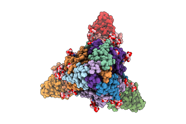
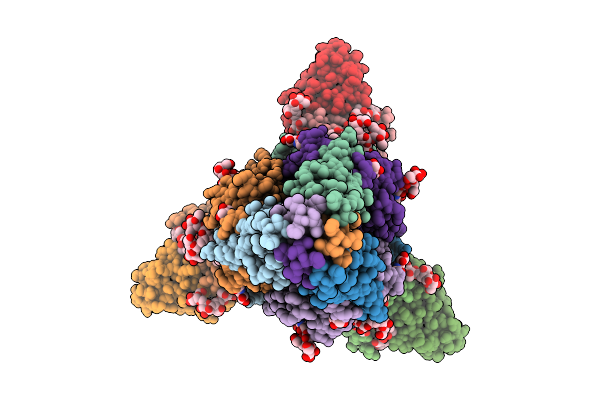
Structure Of The Membrane Soluble Spike Complex From The Lassa Virus In A C3-Symmetric Map
Organism: Lassa virus (strain mouse/sierra leone/josiah/1976)
Method: ELECTRON MICROSCOPY Release Date: 2021-12-29 Classification: VIRAL PROTEIN Ligands: NAG |
 |
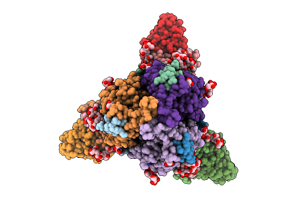
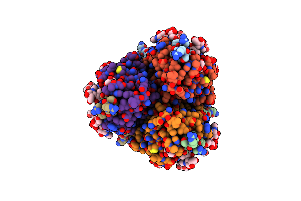
Structure Of The Membrane Soluble Spike Complex From The Lassa Virus In A C1-Symmetric Map Focused On The Ectodomain
Organism: Lassa virus (strain mouse/sierra leone/josiah/1976)
Method: ELECTRON MICROSCOPY Release Date: 2021-12-29 Classification: VIRAL PROTEIN Ligands: NAG |
 |
Organism: Homo sapiens
Method: X-RAY DIFFRACTION Resolution:3.05 Å Release Date: 2020-06-10 Classification: Transferase/DNA Ligands: MG, SAH |
 |
Organism: Homo sapiens
Method: X-RAY DIFFRACTION Resolution:3.00 Å Release Date: 2020-06-10 Classification: Transferase/DNA Ligands: SAH, MG |
 |
Organism: Homo sapiens
Method: X-RAY DIFFRACTION Resolution:2.95 Å Release Date: 2020-06-10 Classification: Transferase/DNA Ligands: SAH, MG |
 |
Organism: Homo sapiens
Method: X-RAY DIFFRACTION Resolution:2.95 Å Release Date: 2020-06-10 Classification: Transferase/DNA Ligands: MG, SAH |
 |
Organism: Homo sapiens
Method: X-RAY DIFFRACTION Resolution:2.11 Å Release Date: 2020-03-04 Classification: Oxidoreductase/Inhibitor Ligands: NAP, CL, PWV, FLC |
 |
Organism: Homo sapiens, Ebola virus
Method: ELECTRON MICROSCOPY Release Date: 2020-02-12 Classification: VIRAL PROTEIN |