
Search Count: 30
 |
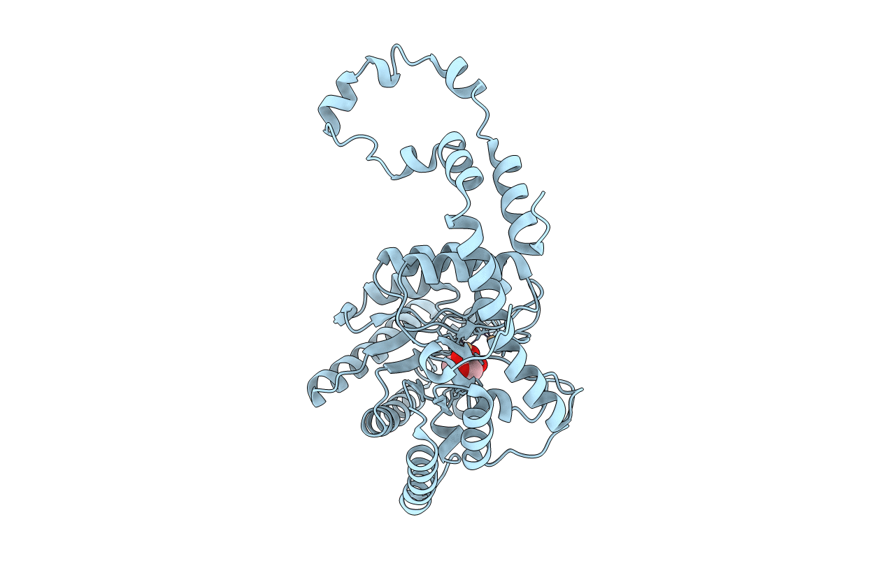
Joint X-Ray And Neutron Structure Of Streptomyces Rubiginosus D-Xylose Isomerase In Complex With Two Cd2+ Ions And Cyclic Beta-L-Arabinose
Organism: Streptomyces rubiginosus
Method: NEUTRON DIFFRACTION, X-RAY DIFFRACTION Resolution:2.00 Å Release Date: 2014-09-03 Classification: ISOMERASE Ligands: CD, ARB, DOD |
 |
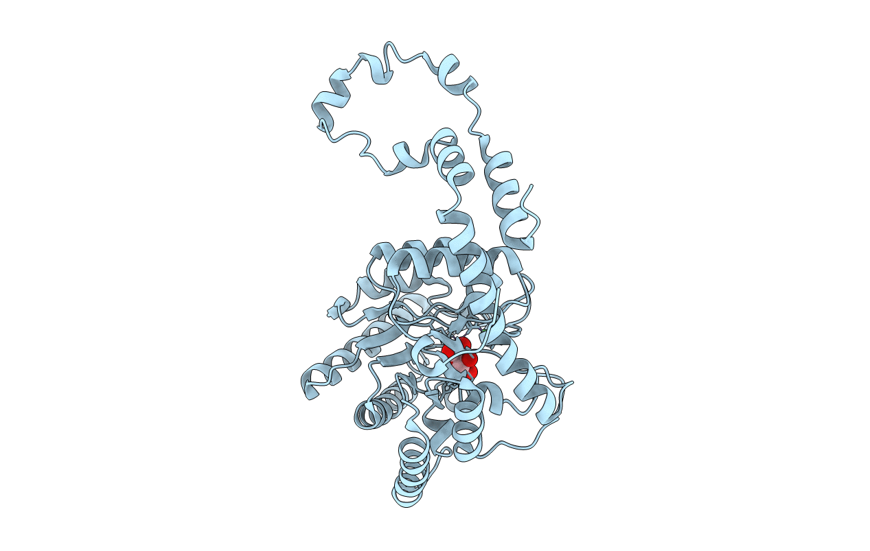
Joint X-Ray And Neutron Structure Of Streptomyces Rubiginosus D-Xylose Isomerase In Complex With Two Ni2+ Ions And Linear L-Arabinose
Organism: Streptomyces rubiginosus
Method: X-RAY DIFFRACTION, NEUTRON DIFFRACTION Resolution:1.80 Å Release Date: 2014-09-03 Classification: ISOMERASE Ligands: NI, LAI, DOD |
 |
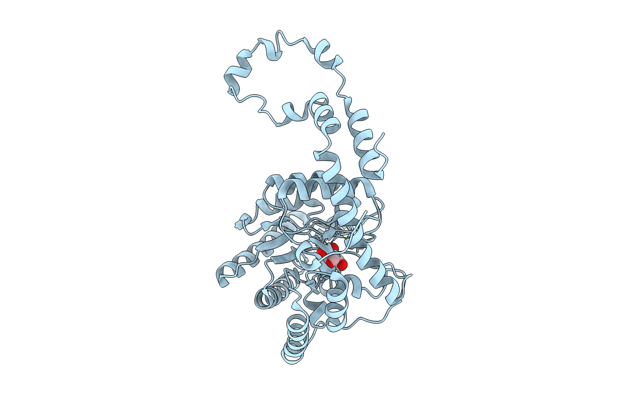
Room Temperature X-Ray Structure Of D-Xylose Isomerase In Complex With Two Cd2+ Ions And L-Ribulose
Organism: Streptomyces rubiginosus
Method: X-RAY DIFFRACTION Resolution:1.55 Å Release Date: 2014-09-03 Classification: ISOMERASE Ligands: CD, RUU |
 |
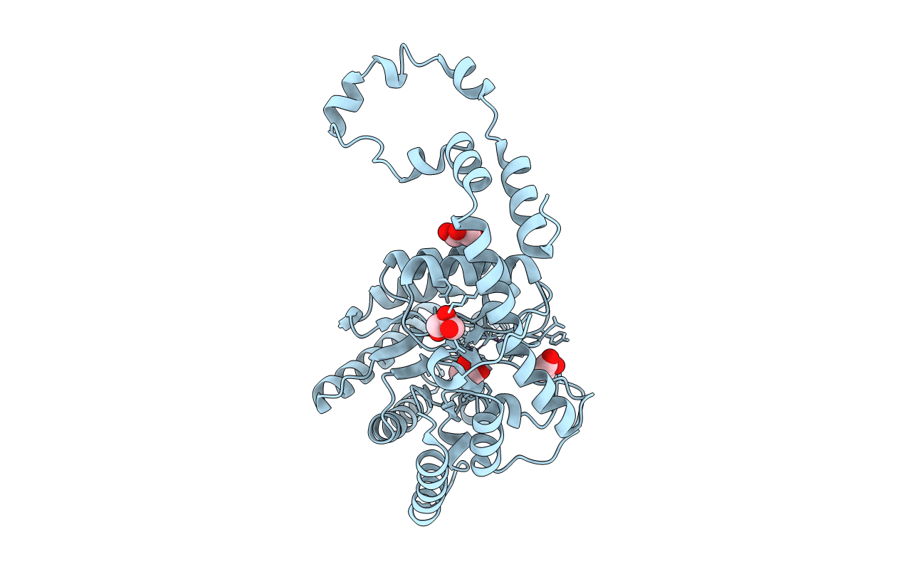
Room Temperature X-Ray Structure Of D-Xylose Isomerase In Complex With Two Ni2+ Ions And L-Ribulose
Organism: Streptomyces rubiginosus
Method: X-RAY DIFFRACTION Resolution:1.70 Å Release Date: 2014-09-03 Classification: ISOMERASE Ligands: NI, 34V |
 |
Room Temperature X-Ray Structure Of D-Xylose Isomerase In Complex With Two Mg2+ Ions And L-Ribulose
Organism: Streptomyces rubiginosus
Method: X-RAY DIFFRACTION Resolution:1.56 Å Release Date: 2014-09-03 Classification: ISOMERASE Ligands: MG, RUU |
 |
Room Temperature X-Ray Structure Of D-Xylose Isomerase In Complex With Two Ni2+ Ions And L-Ribose
Organism: Streptomyces rubiginosus
Method: X-RAY DIFFRACTION Resolution:1.60 Å Release Date: 2014-09-03 Classification: ISOMERASE Ligands: NI, Z6J |
 |
Room Temperature X-Ray Structure Of D-Xylose Isomerase In Complex With Two Mg2+ Ions And L-Ribose
Organism: Streptomyces rubiginosus
Method: X-RAY DIFFRACTION Resolution:1.55 Å Release Date: 2014-09-03 Classification: ISOMERASE Ligands: MG, 32O |
 |
Room Temperature X-Ray Structure Of D-Xylose Isomerase Complexed With 2Cd(2+) Co-Factors And D12-D-Alpha-Glucose In The Cyclic Form
Organism: Streptomyces rubiginosus
Method: X-RAY DIFFRACTION Resolution:2.00 Å Release Date: 2010-06-16 Classification: ISOMERASE Ligands: CD, GLC |
 |
Room Temperature Structure Of D-Xylose Isomerase In Complex With 2Ni(2+) Co-Factors And D12-D-Glucose In The Linear Form
Organism: Streptomyces rubiginosus
Method: X-RAY DIFFRACTION Resolution:1.53 Å Release Date: 2010-06-16 Classification: ISOMERASE Ligands: NI, GLO |
 |
Room Temperature X-Ray Structure Of D-Xylose Isomerase In Complex With 2Cd(2+) Co-Factors
Organism: Streptomyces rubiginosus
Method: X-RAY DIFFRACTION Resolution:1.80 Å Release Date: 2010-06-16 Classification: ISOMERASE Ligands: CD |
 |
Room Temperature Structure Of D-Xylose Isomerase In Complex With 2Ni(2+) Co-Factors
Organism: Streptomyces rubiginosus
Method: X-RAY DIFFRACTION Resolution:1.80 Å Release Date: 2010-06-16 Classification: ISOMERASE Ligands: NI |
 |
Room Temperature X-Ray Mixed-Metal Structure Of D-Xylose Isomerase In Complex With Ni(2+) And Mg(2+) Co-Factors
Organism: Streptomyces rubiginosus
Method: X-RAY DIFFRACTION Resolution:1.60 Å Release Date: 2010-06-16 Classification: ISOMERASE Ligands: NI, MG |
 |
Room Temperature Neutron Structure Of D-Xylose Isomerase In Complex With Two Cd2+ Cations And D12-D-Alpha-Glucose In The Ring Form (Refined Jointly With X-Ray Structure 3Kbm)
Organism: Streptomyces rubiginosus
Method: NEUTRON DIFFRACTION, X-RAY DIFFRACTION Resolution:2.0 Å, 2.0 Å Release Date: 2010-06-16 Classification: ISOMERASE Ligands: CD, GLC, DOD |
 |
Room Temperature Neutron Structure Of D-Xylose Isomerase In Complex With Two Ni2+ Cations And D12-D-Glucose In The Linear Form (Refined Jointly With X-Ray Structure 3Kbn)
Organism: Streptomyces rubiginosus
Method: NEUTRON DIFFRACTION, X-RAY DIFFRACTION Resolution:1.8 Å, 1.53 Å Release Date: 2010-06-16 Classification: ISOMERASE Ligands: NI, GLO, DOD |
 |
Organism: Streptomyces rubiginosus
Method: NEUTRON DIFFRACTION Resolution:2.20 Å Release Date: 2008-08-05 Classification: ISOMERASE Ligands: XUL, MG, OH, DOD |
 |
Organism: Streptomyces rubiginosus
Method: X-RAY DIFFRACTION Resolution:0.94 Å Release Date: 2006-05-16 Classification: ISOMERASE Ligands: MN, GOL |
 |
Organism: Streptomyces rubiginosus
Method: X-RAY DIFFRACTION Resolution:1.80 Å Release Date: 2006-05-16 Classification: ISOMERASE |
 |
Organism: Streptomyces rubiginosus
Method: NEUTRON DIFFRACTION Resolution:2.20 Å Release Date: 2006-05-16 Classification: ISOMERASE Ligands: CO, DOD |
 |
Modes Of Binding Substrates And Their Analogues To The Enzyme D-Xylose Isomerase
Organism: Streptomyces rubiginosus
Method: X-RAY DIFFRACTION Resolution:1.60 Å Release Date: 1994-06-22 Classification: ISOMERASE(INTRAMOLECULAR OXIDOREDUCTASE) Ligands: MN |
 |
Modes Of Binding Substrates And Their Analogues To The Enzyme D-Xylose Isomerase
Organism: Streptomyces rubiginosus
Method: X-RAY DIFFRACTION Resolution:1.60 Å Release Date: 1994-06-22 Classification: ISOMERASE(INTRAMOLECULAR OXIDOREDUCTASE) Ligands: XLS, MN |