
Search Count: 20
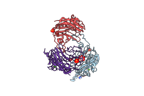 |
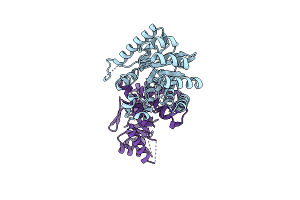
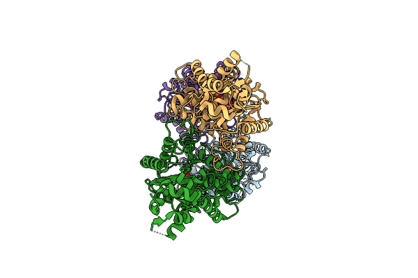
Globular Domain Of The Entamoeba Histolytica Calreticulin In Complex With Glucose
Organism: Entamoeba histolytica hm-1:imss
Method: X-RAY DIFFRACTION Resolution:2.15 Å Release Date: 2016-08-31 Classification: SUGAR BINDING PROTEIN Ligands: CA, SO4, NH4, CL, ACT, BGC |
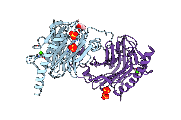 |
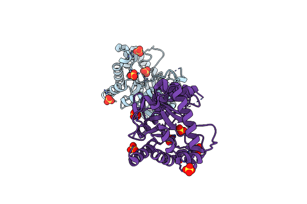
Globular Domain Of The Entamoeba Histolytica Calreticulin In Complex With Glucose
Organism: Entamoeba histolytica hm-1:imss
Method: X-RAY DIFFRACTION Resolution:2.90 Å Release Date: 2016-08-31 Classification: SUGAR BINDING PROTEIN Ligands: SO4, CA, CL, GOL |
 |
Organism: Trypanosoma cruzi (strain cl brener)
Method: X-RAY DIFFRACTION Resolution:2.45 Å Release Date: 2016-08-31 Classification: CHAPERONE Ligands: ACY, CL, BGC |
 |
Organism: Homo sapiens
Method: X-RAY DIFFRACTION Resolution:2.30 Å Release Date: 2016-08-31 Classification: calcium-binding protein Ligands: CA, CL |
 |
Organism: Mus musculus
Method: X-RAY DIFFRACTION Resolution:2.77 Å Release Date: 2013-08-14 Classification: HYDROLASE Ligands: 04A |
 |
Organism: Bacillus anthracis
Method: X-RAY DIFFRACTION Resolution:2.61 Å Release Date: 2012-03-14 Classification: TRANSFERASE Ligands: 78H, SO4 |
 |
Organism: Bacillus anthracis
Method: X-RAY DIFFRACTION Resolution:2.60 Å Release Date: 2012-03-14 Classification: TRANSFERASE Ligands: XHP, PHB, SO4 |
 |
Organism: Bacillus anthracis
Method: X-RAY DIFFRACTION Resolution:2.30 Å Release Date: 2012-03-14 Classification: TRANSFERASE Ligands: XHP, SO4 |
 |
Organism: Bacillus anthracis
Method: X-RAY DIFFRACTION Resolution:2.50 Å Release Date: 2012-03-14 Classification: TRANSFERASE Ligands: POP, XHP, SO4 |
 |
Organism: Bacillus anthracis
Method: X-RAY DIFFRACTION Resolution:2.30 Å Release Date: 2012-03-14 Classification: TRANSFERASE Ligands: XTZ, SO4, XHP, YTZ |
 |
Organism: Yersinia pestis
Method: X-RAY DIFFRACTION Resolution:2.70 Å Release Date: 2012-03-14 Classification: TRANSFERASE Ligands: PT1 |
 |
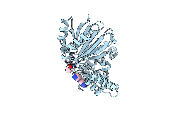
Crystal Structure Of The Yersinia Pestis Dihydropteroate Synthetase With Substrate Transition State Complex.
Organism: Yersinia pestis
Method: X-RAY DIFFRACTION Resolution:2.07 Å Release Date: 2012-03-14 Classification: TRANSFERASE/TRANSFERASE SUBSTRATE Ligands: XHP, POP, PAB, MG |
 |
Crystal Structure Of The Yersinia Pestis Dihydropteroate Synthase With Sulfonamide Drug Complex.
Organism: Yersinia pestis
Method: X-RAY DIFFRACTION Resolution:2.10 Å Release Date: 2012-03-14 Classification: TRANSFERASE/ANTIBIOTIC/INHIBITOR Ligands: HH2, MG, 08D |
 |
Organism: Yersinia pestis
Method: X-RAY DIFFRACTION Resolution:2.08 Å Release Date: 2012-03-14 Classification: TRANSFERASE |
 |
Structural And Mechanistic Studies Of Catalysis And Sulfa Drug Resistance In Dihydropteroate Synthase
Organism: Bacillus anthracis
Method: X-RAY DIFFRACTION Resolution:2.50 Å Release Date: 2012-03-14 Classification: TRANSFERASE Ligands: SO4 |
 |
Organism: Mus musculus
Method: X-RAY DIFFRACTION Resolution:2.42 Å Release Date: 2012-01-11 Classification: HYDROLASE Ligands: CL |
 |
Organism: Mus musculus
Method: X-RAY DIFFRACTION Resolution:2.85 Å Release Date: 2012-01-11 Classification: HYDROLASE Ligands: PO4 |
 |
Organism: Mus musculus
Method: X-RAY DIFFRACTION Resolution:2.80 Å Release Date: 2012-01-11 Classification: HYDROLASE Ligands: GLU |
 |
Organism: Leishmania infantum
Method: X-RAY DIFFRACTION Resolution:1.80 Å Release Date: 2008-01-15 Classification: HYDROLASE Ligands: ZN, SPD, ACY |
 |
Crystal Structure Of The Leishmania Infantum Glyoxalase Ii With D-Lactate At The Active Site
Organism: Leishmania infantum
Method: X-RAY DIFFRACTION Resolution:1.90 Å Release Date: 2008-01-15 Classification: HYDROLASE Ligands: ZN, SPD, LAC |