
Search Count: 15
 |
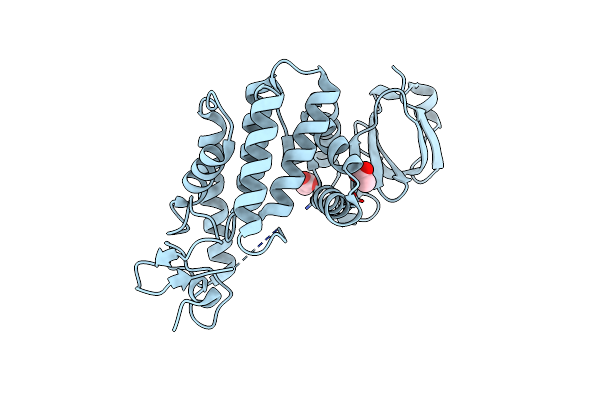
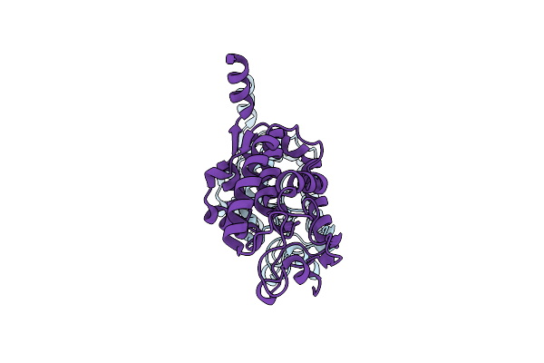
Organism: Saccharomyces cerevisiae
Method: SOLUTION NMR Release Date: 2018-09-12 Classification: MEMBRANE PROTEIN |
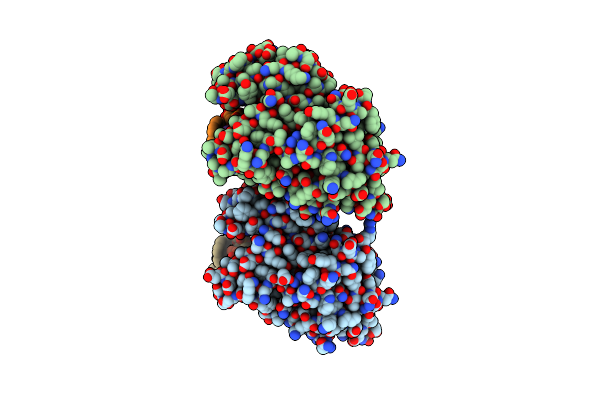 |
Crystal Structure Of Pyrene- And Phenanthrene-Modified Dna In Complex With The Bpuj1 Endonuclease Binding Domain
Organism: Bacillus pumilus, Synthetic construct
Method: X-RAY DIFFRACTION Resolution:2.67 Å Release Date: 2016-08-17 Classification: HYDROLASE |
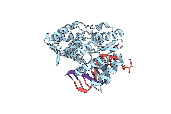 |
Crystal Structure Of Pyrene- And Phenanthrene-Modified Dna In Complex With The Bpuj1 Endonuclease Binding Domain
Organism: Bacillus pumilus, Synthetic construct
Method: X-RAY DIFFRACTION Resolution:1.55 Å Release Date: 2016-08-17 Classification: HYDROLASE |
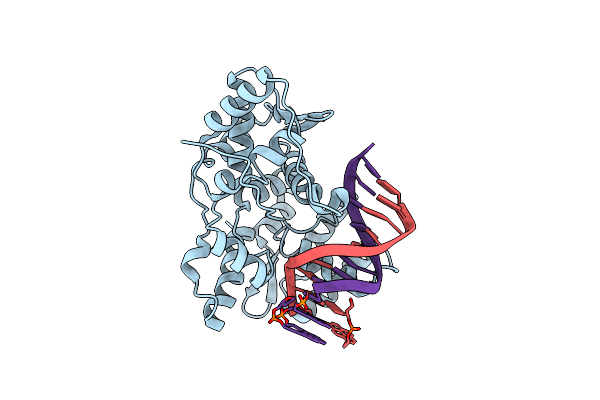 |
Crystal Structure Of Pyrene- And Phenanthrene-Modified Dna In Complex With The Bpuj1 Endonuclease Binding Domain
Organism: Bacillus pumilus, Synthetic construct
Method: X-RAY DIFFRACTION Resolution:1.88 Å Release Date: 2016-08-17 Classification: HYDROLASE |
 |
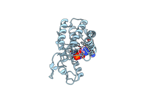
Crystal Structure Of The Fic Domain Of Bep5 Protein (Virb-Translocated Bartonella Effector Protein) From Bartonella Clarridgeiae
Organism: Bartonella clarridgeiae (strain cip 104772 / 73)
Method: X-RAY DIFFRACTION Resolution:2.95 Å Release Date: 2016-01-13 Classification: PROTEIN BINDING Ligands: EDO |
 |
Crystal Structure Of Bepc Protein (Virb-Translocated Bartonella Effector Protein) With Bound Amppnp From Bartonella Tribocorum
Organism: Bartonella tribocorum
Method: X-RAY DIFFRACTION Resolution:1.70 Å Release Date: 2015-10-14 Classification: TRANSFERASE Ligands: ANP, EDO, GLY |
 |
Crystal Structure Of The N-Terminal Fic Domain Of Bep8 Protein (Virb-Translocated Bartonella Effector Protein) From Bartonella Sp. 1-1C
Organism: Bartonella sp. 1-1c
Method: X-RAY DIFFRACTION Release Date: 2015-06-10 Classification: PROTEIN BINDING Ligands: EDO |
 |
Crystal Structure Of Bep1 Protein (Virb-Translocated Bartonella Effector Protein) From Bartonella Clarridgeiae
Organism: Bartonella clarridgeiae
Method: X-RAY DIFFRACTION Resolution:1.90 Å Release Date: 2014-10-08 Classification: CELL ADHESION Ligands: ACT |
 |
Crystal Structure Of The N-Terminal Fic Domain Of A Putative Cell Filamentation Protein (Virb-Translocated Bep Effector Protein) With Bound Adp From Bartonella Quintana
Organism: Bartonella quintana
Method: X-RAY DIFFRACTION Resolution:1.55 Å Release Date: 2014-09-24 Classification: TRANSFERASE Ligands: ADP, MG |
 |
Crystal Structure Of The N-Terminal Fic Domain Of A Putative Cell Filamentation Protein (Virb-Translocated Bep Effector Protein) From Bartonella Quintana
Organism: Bartonella quintana
Method: X-RAY DIFFRACTION Resolution:2.00 Å Release Date: 2014-08-06 Classification: TRANSFERASE Ligands: CL, BR, IOD |
 |
Crystal Structure Of The N-Terminal Fic Domain Of Bartonella Effector Protein (Bep); Substrate Of Virb T4Ss (Virb-Translocated Bep Effector Protein) From Bartonella Sp. Ar 15-3
Organism: Bartonella sp. ar 15-3
Method: X-RAY DIFFRACTION Resolution:1.85 Å Release Date: 2014-08-06 Classification: CELL ADHESION Ligands: EDO |
 |
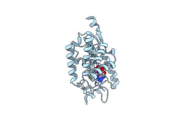
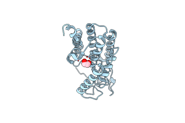
Crystal Structure Of Human Udp-Glucose Dehydrogenase Glu161Gln, In Complex With Thiohemiacetal Intermediate
Organism: Homo sapiens
Method: X-RAY DIFFRACTION Resolution:2.30 Å Release Date: 2009-11-17 Classification: OXIDOREDUCTASE Ligands: UPG, EDO, EPE |
 |
Organism: Homo sapiens
Method: X-RAY DIFFRACTION Resolution:2.40 Å Release Date: 2009-09-15 Classification: OXIDOREDUCTASE Ligands: PG4, EDO |
 |
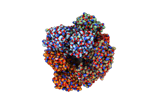
Crystal Structure Of Human Udp-Glucose Dehydrogenase Product Complex With Udp-Glucuronate
Organism: Homo sapiens
Method: X-RAY DIFFRACTION Resolution:2.10 Å Release Date: 2007-07-10 Classification: OXIDOREDUCTASE Ligands: NAD, UGA, CL, EDO |
 |
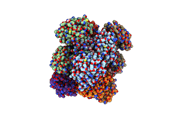
Structure Of Human Udp-Glucose Dehydrogenase Complexed With Nadh And Udp-Glucose
Organism: Homo sapiens
Method: X-RAY DIFFRACTION Resolution:2.00 Å Release Date: 2007-07-03 Classification: OXIDOREDUCTASE Ligands: CL, NAI, UPG |