
Search Count: 8,864
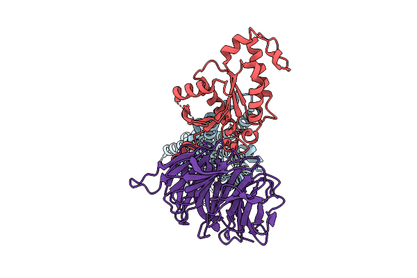 |
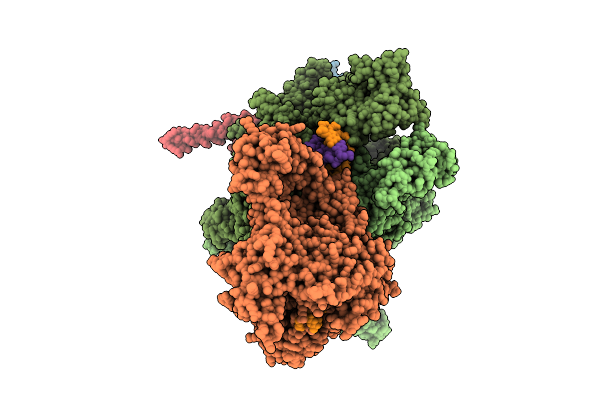
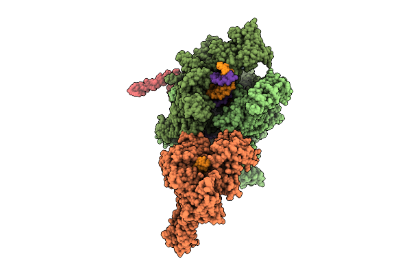
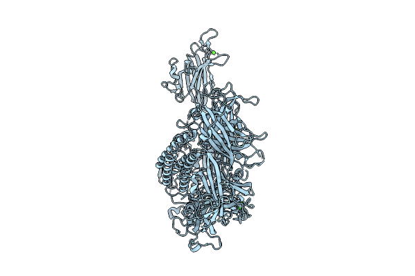
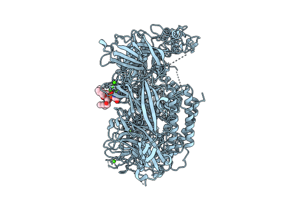
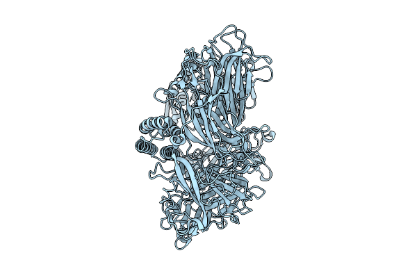
Human P2Ry8 Bound To S-Geranylgeranyl-L-Glutathione In Complex With Minig13
Organism: Homo sapiens
Method: ELECTRON MICROSCOPY Release Date: 2025-11-05 Classification: MEMBRANE PROTEIN Ligands: A1BIG |
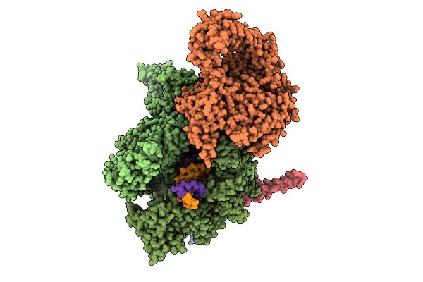 |
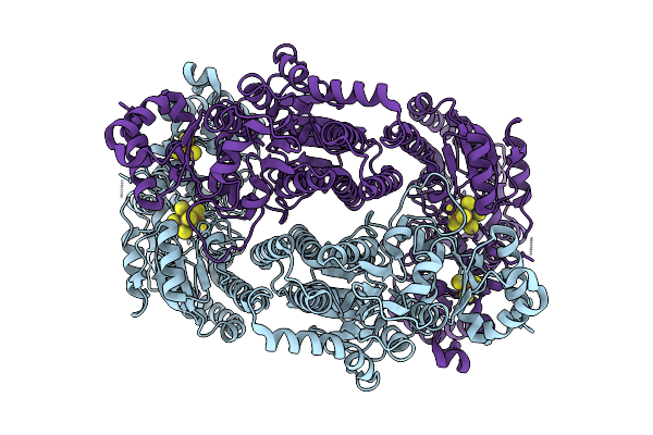
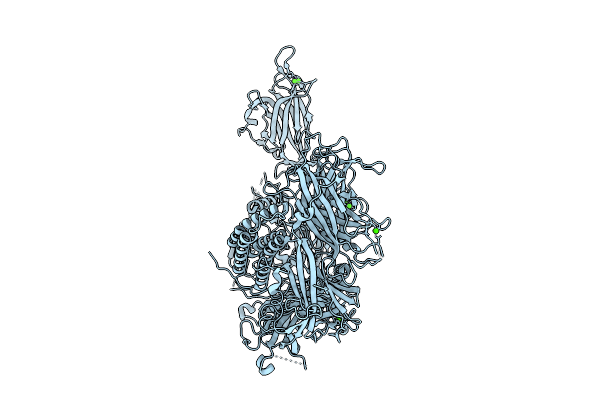
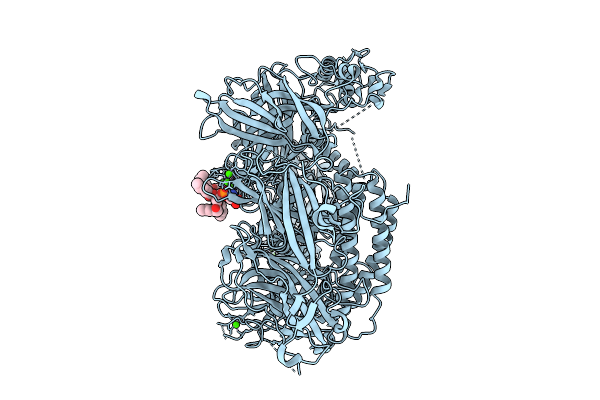
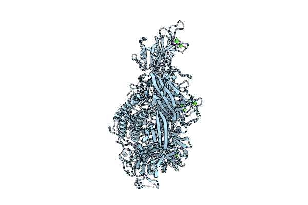
Organism: Escherichia coli, Synthetic construct
Method: ELECTRON MICROSCOPY Release Date: 2025-11-05 Classification: TRANSCRIPTION Ligands: MG, ZN |
 |
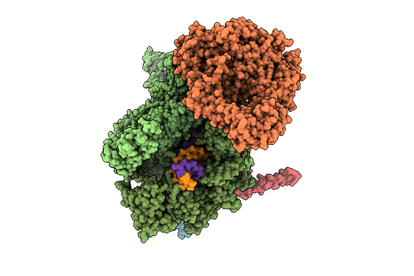
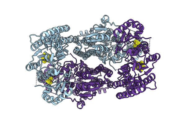
Organism: Escherichia coli, Synthetic construct
Method: ELECTRON MICROSCOPY Release Date: 2025-11-05 Classification: TRANSCRIPTION Ligands: MG, ZN |
 |
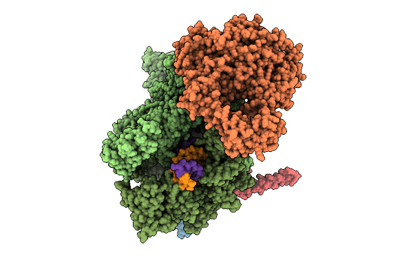
Organism: Escherichia coli, Synthetic construct
Method: ELECTRON MICROSCOPY Release Date: 2025-11-05 Classification: TRANSCRIPTION Ligands: MG, ZN |
 |
Organism: Escherichia coli, Synthetic construct
Method: ELECTRON MICROSCOPY Release Date: 2025-11-05 Classification: TRANSCRIPTION Ligands: MG, ZN |
 |
Organism: Escherichia coli, Synthetic construct
Method: ELECTRON MICROSCOPY Release Date: 2025-11-05 Classification: TRANSCRIPTION Ligands: MG, ZN |
 |
Organism: Escherichia coli, Synthetic construct
Method: ELECTRON MICROSCOPY Release Date: 2025-11-05 Classification: TRANSCRIPTION Ligands: MG, ZN |
 |
Organism: Geobacter metallireducens
Method: ELECTRON MICROSCOPY Release Date: 2025-11-05 Classification: METAL BINDING PROTEIN Ligands: SF4, S5Q |
 |
Organism: Geobacter metallireducens
Method: ELECTRON MICROSCOPY Release Date: 2025-11-05 Classification: METAL BINDING PROTEIN Ligands: SF4, S5Q |
 |
Mouse Otoferlin (216-1931) In The Lipid-Free, Ca2+-Bound State, "Open" Conformation (Class 2)
Organism: Mus musculus
Method: ELECTRON MICROSCOPY Release Date: 2025-11-05 Classification: MEMBRANE PROTEIN Ligands: CA |
 |
Mouse Otoferlin (216-1931) In Complex With A Lipid Nanodisc (Comprising 25% Ps And 5% Pip2)
Organism: Mus musculus
Method: ELECTRON MICROSCOPY Release Date: 2025-11-05 Classification: MEMBRANE PROTEIN Ligands: CA, PSF |
 |
Mouse Otoferlin (216-1931) In The Lipid-Free Ca2+-Bound State, "Open" Conformation (Class 1)
Organism: Mus musculus
Method: ELECTRON MICROSCOPY Release Date: 2025-11-05 Classification: MEMBRANE PROTEIN Ligands: CA |
 |
Mouse Otoferlin (Residues 216-1931) In The Lipid-Bound State (Merged Datasets)
Organism: Mus musculus
Method: ELECTRON MICROSCOPY Release Date: 2025-11-05 Classification: MEMBRANE PROTEIN Ligands: CA, PSF |
 |
Mouse Otoferlin (216-1931) In The Lipid-Free, Ca2+-Free State ("Loose" Conformation)
Organism: Mus musculus
Method: ELECTRON MICROSCOPY Release Date: 2025-11-05 Classification: MEMBRANE PROTEIN |
 |
Mouse Otoferlin (216-1931) In The Lipid-Free Ca2+-Bound State, "Closed-Like" Conformation
Organism: Mus musculus
Method: ELECTRON MICROSCOPY Release Date: 2025-11-05 Classification: MEMBRANE PROTEIN Ligands: CA |
 |
Crystal Structure Of The Mycobacterium Tuberculosis Regulator Virs (N-Terminal Fragment 4-208) In Complex With The Drug Candidate Alpibectir
Organism: Mycobacterium tuberculosis h37rv
Method: X-RAY DIFFRACTION Release Date: 2025-10-29 Classification: TRANSCRIPTION Ligands: YPQ |
 |
Gag Ca-Sp1 (T8I) Immature Lattice Bound With Bevirimat From Enveloped Virus Like Particles
Organism: Human immunodeficiency virus type 1 (hbx2 isolate)
Method: ELECTRON MICROSCOPY Release Date: 2025-10-29 Classification: VIRAL PROTEIN Ligands: 2I4, IHP |
 |
Structure Of Thioferritin (Pfdpsl) With Ferrihydrite Growth At A Single Three-Fold Pore.
Organism: Pyrococcus furiosus
Method: ELECTRON MICROSCOPY Release Date: 2025-10-29 Classification: METAL BINDING PROTEIN Ligands: FE, OXY, O |
 |
Structure Of Cathepsin B1 From Schistosoma Mansoni (Smcb1) In Complex With A Carborane Inhibitor
Organism: Schistosoma mansoni
Method: X-RAY DIFFRACTION Release Date: 2025-10-29 Classification: HYDROLASE Ligands: A1IHQ, EDO |
 |
Organism: Homo sapiens, Lama glama
Method: X-RAY DIFFRACTION Release Date: 2025-10-29 Classification: PROTEIN TRANSPORT |